Jigsaw Puzzle : False-col SEM of single neurone of cerebral cortex


![]()
Jigsaw Puzzles from Science Photo Library
False-col SEM of single neurone of cerebral cortex
False-colour scanning electron micrograph (SEM) of a single neurone (nerve cell) of the human cerebral cortex - the outer grey matter of the brain. Neurones exist in varying sizes & shapes throughout the nervous system. A basic neurone consists of a cell body with processes of two types: a single axon (the output, effector nerve fibre) and one or more dendrites, smaller, input processes that serve as sensory receptors. In the cerebral cortex, neurones of comparable structure are arranged in a variable number of layers. Magnification: x3, 100 at 6x7cm, x1, 550 at 35mm size
Science Photo Library features Science and Medical images including photos and illustrations
Media ID 6449233
© CNRI/SCIENCE PHOTO LIBRARY PHOTO LIBRARY
Cerebral Cortex Grey Matter Nerve Cell Nervous Neurone System Cells
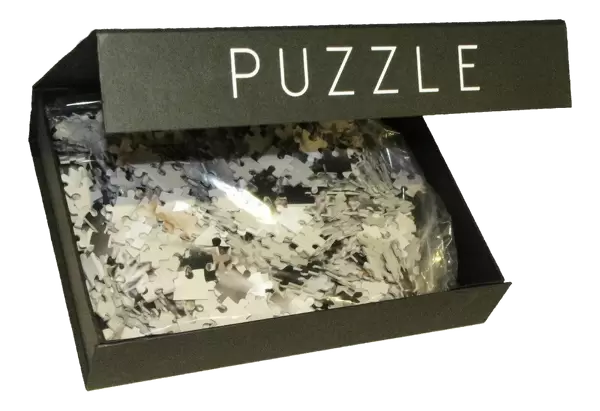
Jigsaw Puzzle (500 Pieces)
Discover the intricacies of the human brain with our Media Storehouse Jigsaw Puzzles. This scientific masterpiece, featuring a False-colour SEM of a single neurone of the cerebral cortex from Science Photo Library, offers an engaging and educational puzzle experience. Challenge yourself and your family to piece together this intricate image of the outer grey matter of the brain, revealing the complex structure of a single neurone. A captivating addition to your home or classroom, this puzzle encourages critical thinking and problem-solving skills, while providing a glimpse into the fascinating world of neuroscience.
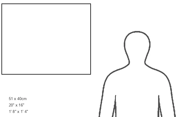
500 piece puzzles are custom made in Australia and hand-finished on 100% recycled 1.6mm thick laminated puzzle boards. There is a level of repetition in jigsaw shapes with each matching piece away from its pair. The completed puzzle measures 40x51cm and is delivered packaged in an attractive presentation box specially designed to fit most mail slots with a unique magnetic lid
Jigsaw Puzzles are an ideal gift for any occasion
Estimated Product Size is 50.7cm x 40.3cm (20" x 15.9")
These are individually made so all sizes are approximate
Artwork printed orientated as per the preview above, with landscape (horizontal) or portrait (vertical) orientation to match the source image.
EDITORS COMMENTS
This false-colour scanning electron micrograph (SEM) offers a mesmerizing glimpse into the intricate world of a single neurone found in the cerebral cortex, the outer grey matter of the human brain. Neurones, or nerve cells, come in various sizes and shapes throughout our nervous system, playing a vital role in transmitting electrical signals. The image showcases a basic neurone structure consisting of a cell body with two distinct types of processes. The first is an axon, serving as an output or effector nerve fibre responsible for relaying information to other cells. The second type is dendrites, smaller input processes acting as sensory receptors that receive signals from other neurones. In the cerebral cortex specifically, these remarkable neurones are organized into different layers with comparable structures but varying numbers. This arrangement contributes to the complex functioning and processing capabilities of this critical region within our brains. With its impressive magnification power at x3,100 on a 6x7cm print and x1,550 on 35mm size prints, this SEM allows us to appreciate the intricate details and delicate beauty inherent in our own neural architecture. It serves as a reminder of just how awe-inspiring and sophisticated our nervous system truly is.
MADE IN AUSTRALIA
Safe Shipping with 30 Day Money Back Guarantee
FREE PERSONALISATION*
We are proud to offer a range of customisation features including Personalised Captions, Color Filters and Picture Zoom Tools
SECURE PAYMENTS
We happily accept a wide range of payment options so you can pay for the things you need in the way that is most convenient for you
* Options may vary by product and licensing agreement. Zoomed Pictures can be adjusted in the Cart.