Fraud Blocker Skip to main content

Immune Response Collection

"Unleashing the Power of Immune Response: A Battle against Disease" In the microscopic world

Background imageImmune Response Collection: T lymphocytes and cancer cell, SEM C001 / 1679

T lymphocytes and cancer cell, SEM C001 / 1679
T lymphocytes and cancer cell. Coloured scanning electron micrograph (SEM) of T lymphocyte cells (green) attached to a cancer cell

Background imageImmune Response Collection: Neutrophil engulfing MRSA, SEM C018 / 8596

Neutrophil engulfing MRSA, SEM C018 / 8596
Neutrophil engulfing MRSA. Coloured scanning electron micrograph (SEM) of a neutrophil white blood cell (green) engulfing methicillin-resistant Staphylococcus aureus bacteria (MRSA, pink)

Background imageImmune Response Collection: Bacteria infecting a macrophage, SEM

Bacteria infecting a macrophage, SEM
Bacteria infecting a macrophage. Coloured scanning electron micrograph (SEM) of Mycobacterium tuberculosis bacteria (purple) infecting a macrophage white blood cell

Background imageImmune Response Collection: Phagocytosis of fungal spores, SEM

Phagocytosis of fungal spores, SEM
Phagocytosis of a fungus spore. Coloured scanning electron micrograph (SEM) of a neutrophil (blue) phagocytosing (engulfing and destroying) spores from the fungus Aspergillus fumigatus (red)

Background imageImmune Response Collection: Bacteria infecting a macrophage, SEM

Bacteria infecting a macrophage, SEM
Bacteria infecting a macrophage. Coloured scanning electron micrograph (SEM) of Mycobacterium tuberculosis bacteria (purple) infecting a macrophage white blood cell

Background imageImmune Response Collection: Rhinovirus and antibody, molecular model C015 / 7139

Rhinovirus and antibody, molecular model C015 / 7139
Rhinovirus. Molecular model of the antigen-binding fragment (Fab) from a strongly neutralising antibody bound to a human rhinovirus 14 (HRV-14) particle

Background imageImmune Response Collection: Phagocytosis of fungus spores, SEM

Phagocytosis of fungus spores, SEM
Phagocytosis of fungus spores. Coloured scanning electron micrograph (SEM) of spores from the fungus Aspergillus fumigatus (red) being phagocytosed (engulfed and destroyed) by a dendritic cell (brown)

Background imageImmune Response Collection: Foot-and-mouth disease virus F006 / 9556

Foot-and-mouth disease virus F006 / 9556
Foot-and-mouth disease virus. Molecular model of the foot-and-mouth disease (FMD) virus (Aphtae epizooticae) protein coat (capsid)

Background imageImmune Response Collection: Macrophage and lymphocytes, TEM

Macrophage and lymphocytes, TEM
Macrophage and lymphocytes, transmission electron micrograph (TEM). Macrophage surrounded by lymphocytes in a lymph node. This view includes many intercellular contacts

Background imageImmune Response Collection: Rhinovirus and antibody, molecular model C015 / 7138

Rhinovirus and antibody, molecular model C015 / 7138
Rhinovirus. Molecular model of the antigen-binding fragment (Fab) from a strongly neutralising antibody bound to a human rhinovirus 14 (HRV-14) particle

Background imageImmune Response Collection: Murine norovirus with antibody fragments

Murine norovirus with antibody fragments
Murine norovirus (MNV) with antibody fragments, computer model. This image was created using molecular modelling software and data from cryo- electron microscopy

Background imageImmune Response Collection: Cutaway of Lymph Node with each Tissue type highlighted

Cutaway of Lymph Node with each Tissue type highlighted

Background imageImmune Response Collection: Haemagglutinin viral surface protein F007 / 9932

Haemagglutinin viral surface protein F007 / 9932
Haemagglutinin viral surface protein. Molecular model of haemagglutinin, a surface protein from the influenza virus, complexed with a neutralising antibody

Background imageImmune Response Collection: Haemagglutinin viral surface protein F007 / 9931

Haemagglutinin viral surface protein F007 / 9931
Haemagglutinin viral surface protein. Molecular model of haemagglutinin, a surface protein from the influenza virus, complexed with a neutralising antibody

Background imageImmune Response Collection: Neutrophil engulfing MRSA, SEM C018 / 8597

Neutrophil engulfing MRSA, SEM C018 / 8597
Neutrophil engulfing MRSA. Coloured scanning electron micrograph (SEM) of a neutrophil white blood cell (bottom) engulfing methicillin-resistant Staphylococcus aureus bacteria (MRSA, yellow)

Background imageImmune Response Collection: Human antitumour antibody molecule

Human antitumour antibody molecule
Human antitumour antibody. Molecular model showing the antitumour antibody BR96 complexed with part of the Lewis antigen. The Lewis antigen is expressed on the surface of human carcinoma cells

Background imageImmune Response Collection: Toll-like receptor 3 and RNA F006 / 9666

Toll-like receptor 3 and RNA F006 / 9666
Toll-like receptor 3 and RNA. Molecular model of the toll-like receptor 3 (TLR3) protein (pink and blue) bound to a strand of RNA (ribonucleic acid, green and yellow)

Background imageImmune Response Collection: Interferon regulatory factor molecule F006 / 9630

Interferon regulatory factor molecule F006 / 9630
Interferon regulatory factor. Molecular model of interferon regulatory factor 3 (IRF3, coils at right and left) bound to a DNA (deoxyribonucleic acid) molecule (red and blue)

Background imageImmune Response Collection: H5N1 Haemagglutinin protein subunit F006 / 9590

H5N1 Haemagglutinin protein subunit F006 / 9590
H5N1 haemagglutinin protein subunit. Molecular model of the haemagglutinin HA(5) subunit. Haemagglutinin is a surface protein from the influenza A virus

Background imageImmune Response Collection: Haemagglutinin protein subunit F006 / 9479

Haemagglutinin protein subunit F006 / 9479
Haemagglutinin protein subunit. Molecular model of the ectodomain of the haemagglutinin HA(2) subunit. Haemagglutinin is a surface protein from the influenza A virus

Background imageImmune Response Collection: Foot-and-mouth disease virus F006 / 9471

Foot-and-mouth disease virus F006 / 9471
Foot-and-mouth disease virus. Molecular model of the foot-and-mouth disease (FMD) virus Aphtae epizooticae, with antibodies (immunoglobulins)

Background imageImmune Response Collection: Haemagglutinin viral surface protein F006 / 9470

Haemagglutinin viral surface protein F006 / 9470
Haemagglutinin viral surface protein. Molecular model of haemagglutinin, a surface protein from the influenza virus, complexed with a neutralising antibody

Background imageImmune Response Collection: Flu virus surface protein and antibody F006 / 9428

Flu virus surface protein and antibody F006 / 9428
Flu virus surface protein and antibody. Molecular model of the neuraminidase glycoprotein enzyme found on the surface of the influenza (flu) virus

Background imageImmune Response Collection: Interleukin-1 beta molecule F006 / 9369

Interleukin-1 beta molecule F006 / 9369
Interleukin-1 beta, molecular model. This cytokine immune protein is an important mediator of the inflammatory response

Background imageImmune Response Collection: MHC protein-antigen complex F006 / 9363

MHC protein-antigen complex F006 / 9363
MHC protein-antigen complex. Molecular model of the human class I MHC (major histocompatibility complex) protein HLA-B27 complexed with beta-2 microglobulin

Background imageImmune Response Collection: MHC protein complexed with flu virus F006 / 9294

MHC protein complexed with flu virus F006 / 9294
MHC protein complexed with flu virus. Molecular model showing human class II MHC (major histocompatibility complex) protein HLA-DR1 complexed with an influenza (flu) virus peptide

Background imageImmune Response Collection: Antibodies and viruses, artwork

Antibodies and viruses, artwork
Antibodies and viruses. Computer artwork showing antibody, or immunoglobulin, molecules (blue) surrounding virus particles (virions, yellow)

Background imageImmune Response Collection: T-cell receptor bound to enterotoxin

T-cell receptor bound to enterotoxin, molecular model. The T cell receptor (TCR) is a protein complex found on the surface of a type of white blood cell called T lymphocytes (or T cells)

Background imageImmune Response Collection: NK cells attacking cancer cells, artwork

NK cells attacking cancer cells, artwork
NK cells attacking cancer cells. Computer artwork of human Natural Killer (NK) cells (small, round) attacking cancer cells (large, round)

Background imageImmune Response Collection: Immunotherapy, artwork

Immunotherapy, artwork
Immunotherapy. Computer artwork of immunotherapy being used to destroy a tumour (right). Immunotherapy uses the bodys immune system to fight a disease

Background imageImmune Response Collection: Antibodies and bacteria, artwork

Antibodies and bacteria, artwork
Antibodies and bacteria. Computer artwork showing white blood cells (large, round) and antibody, or immunoglobulin, molecules (red) surrounding bacteria (green)

Background imageImmune Response Collection: MHC protein-antigen complex C015 / 1924

MHC protein-antigen complex C015 / 1924
MHC protein-antigen complex. Molecular model of the human class I MHC (major histocompatibility complex) protein HLA-D27 complexed with a peptide antigen

Background imageImmune Response Collection: Mast cells, light micrograph

Mast cells, light micrograph
Mast cells. Light micrograph of several mast cells (pink rounded objects). These leucocytes (white blood cells) contain the chemical mediators histamine, serotonin and heparin

Background imageImmune Response Collection: Granulocyte white blood cell, TEM

Granulocyte white blood cell, TEM
Granulocyte white blood cell. Coloured transmission electron micrograph (TEM) of a neutrophil granulocyte, a type of white blood cell

Background imageImmune Response Collection: Haemagglutinin viral surface protein C015 / 9965

Haemagglutinin viral surface protein C015 / 9965
Haemagglutinin viral surface protein. Molecular model of haemagglutinin, a surface protein from the influenza virus, complexed with a neutralising antibody

Background imageImmune Response Collection: Haemagglutinin viral surface protein C015 / 7124

Haemagglutinin viral surface protein C015 / 7124
Haemagglutinin viral surface protein. Molecular model of haemagglutinin, a surface protein from the influenza virus, complexed with a neutralising antibody

Background imageImmune Response Collection: Haemagglutinin viral surface protein C015 / 9974

Haemagglutinin viral surface protein C015 / 9974
Haemagglutinin viral surface protein. Molecular model of haemagglutinin, a surface protein from the influenza virus, complexed with a neutralising antibody

Background imageImmune Response Collection: Haemagglutinin viral surface protein C015 / 7123

Haemagglutinin viral surface protein C015 / 7123
Haemagglutinin viral surface protein. Molecular model of haemagglutinin, a surface protein from the influenza virus, complexed with a neutralising antibody

Background imageImmune Response Collection: Lung silicosis, light micrograph C015 / 7129

Lung silicosis, light micrograph C015 / 7129
Lung silicosis. Light micrograph of a section through a lung with silicosis, a type of pneumoconiosis caused by exposure to silica dusts. The lung tissue has become fibrous (centre)

Background imageImmune Response Collection: Neutrophil white blood cell, illustration C018 / 0741

Neutrophil white blood cell, illustration C018 / 0741
Neutrophil white blood cell, illustration. Neutrophils are the most abundant white blood cell in humans. They have a multi-lobed nucleus (dark purple)

Background imageImmune Response Collection: Human immune response molecule complex

Human immune response molecule complex. Molecular model showing a human T-cell receptor and an HLA-A leukocyte (white blood cell) antigen bound to a TAX peptide from a virus

Background imageImmune Response Collection: Microglial white blood cell, SEM C019 / 0247

Microglial white blood cell, SEM C019 / 0247
Microglial white blood cell, SEM

Background imageImmune Response Collection: Microglial white blood cell, SEM C019 / 0246

Microglial white blood cell, SEM C019 / 0246
Microglia. Coloured scanning electron micrograph (SEM) of an activated microglial white blood cell. This immune system cell is found in the brain and spinal cord

Background imageImmune Response Collection: Microglial white blood cell, SEM C019 / 0245

Microglial white blood cell, SEM C019 / 0245
Microglia. Coloured scanning electron micrograph (SEM) of an activated microglial white blood cell. This immune system cell is found in the brain and spinal cord

Background imageImmune Response Collection: Human immune response molecule complex C014 / 0871

Human immune response molecule complex C014 / 0871
Human immune response molecule complex. Molecular model showing a human T-cell receptor and an HLA-A leukocyte (white blood cell) antigen bound to a TAX peptide from a virus

Background imageImmune Response Collection: MHC protein-antigen complex

MHC protein-antigen complex. Computer model showing a histocompatibility antigen complexed to a class II MHC (major histocompatibility complex) protein molecule

Background imageImmune Response Collection: Neutrophil engulfing MRSA, SEM C017 / 8301

Neutrophil engulfing MRSA, SEM C017 / 8301
Neutrophil engulfing MRSA. Coloured scanning electron micrograph (SEM) of a neutrophil white blood cell (purple) engulfing methicillin-resistant Staphylococcus aureus bacteria (MRSA, yellow)

Background imageImmune Response Collection: Immune response

Immune response. Fluorescent light micrograph of T lymphocytes (red) surrounding glial cells. Glial cells are support cells for neurons (nerve cells)


All Professionally Made to Order for Quick Shipping

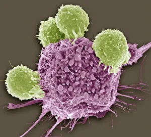
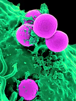
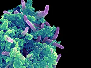
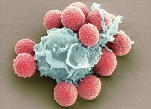
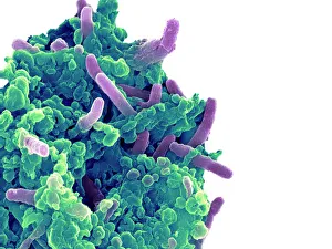
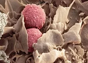
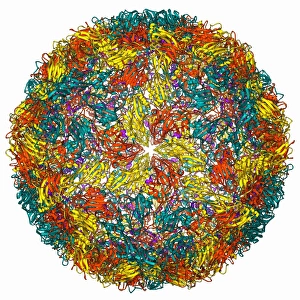
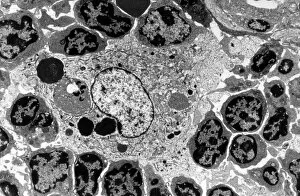
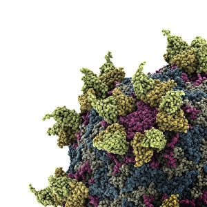
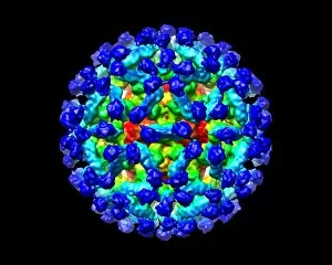
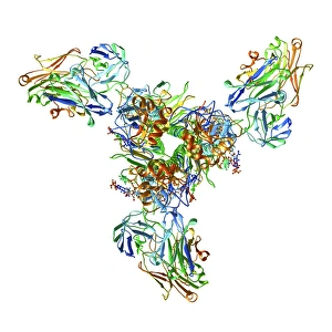
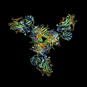
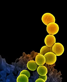
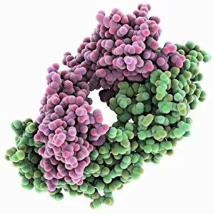
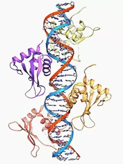
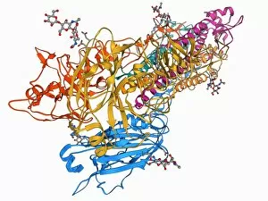
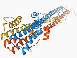
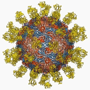
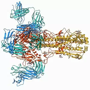
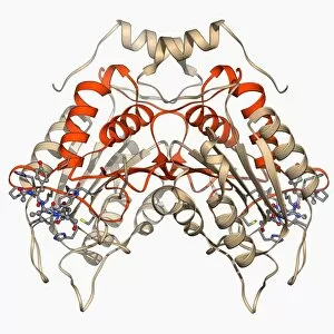
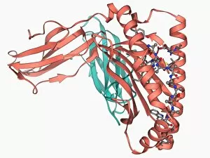
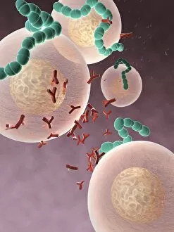
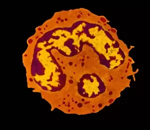
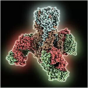
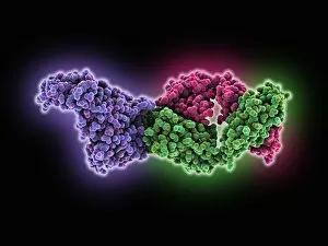
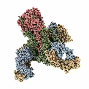
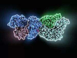
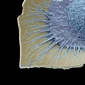
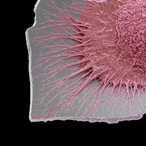
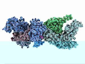
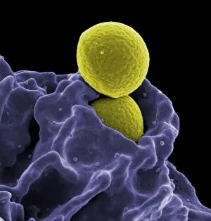
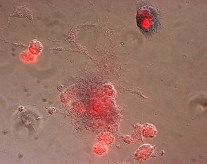
"Unleashing the Power of Immune Response: A Battle against Disease" In the microscopic world, a remarkable defense mechanism unfolds as T lymphocytes take on cancer cells, depicted in stunning detail by SEM C001 / 1679. These vigilant warriors tirelessly seek out and destroy malignant invaders, offering hope in the fight against this devastating disease. Meanwhile, another fierce warrior emerges as a neutrophil engulfs MRSA bacteria with astonishing precision, captured magnificently through SEM C018 / 8596. This powerful immune cell demonstrates its ability to neutralize dangerous pathogens that threaten our well-being. However, not all battles end swiftly. Bacteria infecting macrophages is an ongoing struggle showcased by striking SEM imagery. Witness these tenacious microbes infiltrating and exploiting host cells while our immune system fights back relentlessly. Phagocytosis takes center stage once again as fungal spores fall victim to its gripping power under the watchful eye of SEM technology. The intricate process of engulfment reveals how our body's defenders eliminate potential threats before they can wreak havoc within us. The war rages on with rhinovirus attempting to invade our respiratory system only to be met head-on by antibodies represented in molecular model C015 / 7139. This captivating image showcases the intricate dance between pathogen and defender at a molecular level - a testament to nature's complexity. As we delve deeper into this microscopic realm, phagocytosis reappears vividly capturing fungus spores being devoured under SEM scrutiny. Our immune arsenal leaves no stone unturned when it comes to safeguarding us from harm. Foot-and-mouth disease virus F006 / 9556 serves as yet another reminder of the constant battle fought within us daily. However formidable these adversaries may seem, our immune response stands ready to confront them head-on with unwavering determination. A glimpse into TEM imagery reveals macrophages collaborating harmoniously with lymphocytes, forming an unbreakable alliance against invading pathogens.