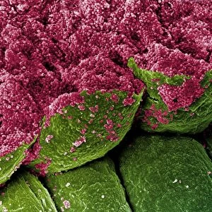
Skin section, SEM

![]()
Wall Art and Photo Gifts from Science Photo Library
Skin section, SEM
Skin section. Coloured scanning electron micrograph (SEM) of a section through human skin. A hair (blue) is protruding through the surface. The top layer of the epidermis is the stratum corneum (cornified layer, light brown). It is comprised of dead, keratinised flattened skin cells, which are continuously sloughed off and replaced with new cells from below. The living layer of the epidermis (dark brown) is called the Malpighian layer. This layer contains cells called melanocytes, which produce the pigment melanin. This pigment darkens the skin when it is exposed to sunlight, producing a suntan. Magnification: x55 when printed 10 centimetres wide
Science Photo Library features Science and Medical images including photos and illustrations
Media ID 6455167
© STEVE GSCHMEISSNER/SCIENCE PHOTO LIBRARY
Epidermal Epidermis False Colour Hair Integument Keratinised Magnified Image Melanin Melanocyte Microscopic Photos Pigment Shaft Skin Stratum Corneum Subjects Cells False Coloured
MADE IN AUSTRALIA
Safe Shipping with 30 Day Money Back Guarantee
FREE PERSONALISATION*
We are proud to offer a range of customisation features including Personalised Captions, Color Filters and Picture Zoom Tools
SECURE PAYMENTS
We happily accept a wide range of payment options so you can pay for the things you need in the way that is most convenient for you
* Options may vary by product and licensing agreement. Zoomed Pictures can be adjusted in the Cart.