
Tumour Collection
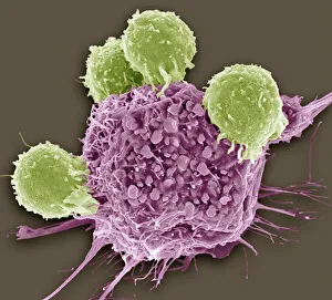
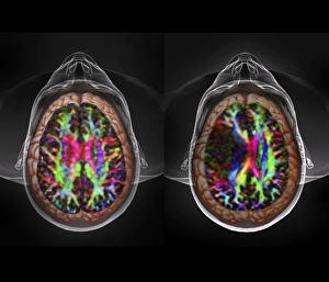
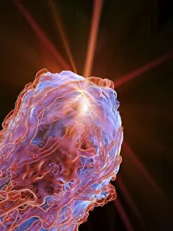
"Tumour: Unveiling the Battle Within" In a microscopic realm, T lymphocytes engage in an intense combat against cancer cells, as captured by SEM C001 / 1679


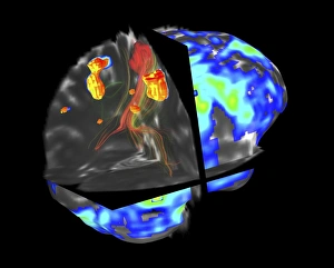
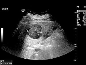
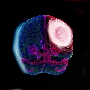


All Professionally Made to Order for Quick Shipping
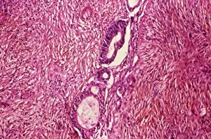
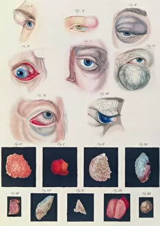
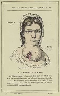
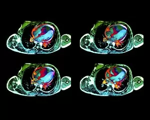
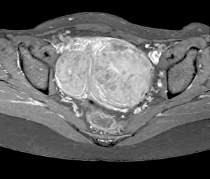
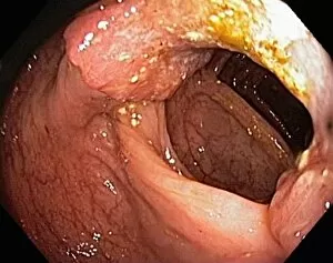
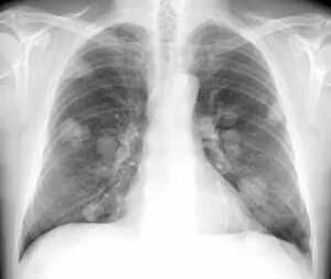
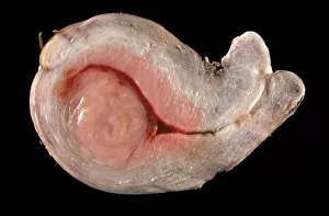
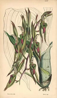
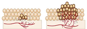
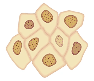
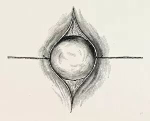
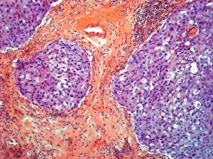
"Tumour: Unveiling the Battle Within" In a microscopic realm, T lymphocytes engage in an intense combat against cancer cells, as captured by SEM C001 / 1679. These brave warriors of our immune system tirelessly fight to protect us from the clutches of disease. Moving to a different battleground, ovarian cancer reveals its intricate nature under the lens of a light micrograph (C015 / 7103). This visual exploration sheds light on the complexity and urgency surrounding this formidable adversary. Amidst these scientific endeavors, nanorobots emerge as potential allies in our war against cancer. Witness their futuristic assault on malignant cells, offering hope for a brighter future where tumors are eradicated (Nanorobot attacking cancer). Beyond the realm of science lies another facet of tumours - congenital eye anomalies. Klinische Darstellungen der provides insight into these mysterious conditions that affect vision and challenge medical professionals' understanding. Exploring further, we encounter "Kyste sereux cloisonne du cou" and "Lipome de la tete, " captivating colour lithographs that depict unique manifestations of tumours in unexpected places. These artistic representations remind us that even within chaos lies beauty waiting to be discovered. Delving into history's archives brings forth Joanna Southcott, an extraordinary fanatic whose beliefs stirred controversy during her time. Like a metaphorical tumour challenging societal norms, she left an indelible mark on history (Joanna Southcott: An Extraordinary Fanatic, 1822). Artistic expressions also capture the essence of tumours; J-L David's masterpiece Chevignard portrays emotions intertwined with physical affliction caused by these growths. Through art's lens emerges empathy for those affected by such ailments (J-L David / Chevignard). Nature too holds its own share of peculiarities – behold Catasetum callosum with its tumor-lipped or callous appearance.