Cushion : Insect microscopy, 19th century


![]()
Home Decor from Science Photo Library
Insect microscopy, 19th century
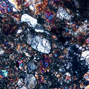
Insect microscopy. 19th century artwork showing microscopic details of insects, as seen under a microscope. An aphid is seen at top centre, with a tsetse fly proboscis immediately below it. Below and left of the proboscis is a feathery type of antennae, as often seen on moths. Claws, as seen on an insects foot, are seen below the feathery antenna and at right of the proboscis. Other items seen include insect larvae and eggs. Artwork taken from the 1883 edition of Jabez Hoggs book The Microscope: its History, Construction and Application
Science Photo Library features Science and Medical images including photos and illustrations
Media ID 6419706
© SCIENCE PHOTO LIBRARY
1883 Antenna Antennae Antennas Aphid Book Claws Construction Drawing Eggs Entomological Foot Larva Larvae Legs Microscopic Microscopy Proboscis Specimen Specimens Text Book
Cushion
Refresh your home decor with a beautiful full photo 16"x16" (40x40cm) cushion, complete with cushion pad insert. Printed on both sides and made from 100% polyester with a zipper on the bottom back edge of the cushion cover. Care Instructions: Warm machine wash, do not bleach, do not tumble dry. Warm iron inside out. Do not dry clean.
Accessorise your space with decorative, soft cushions
Estimated Product Size is 40cm x 40cm (15.7" x 15.7")
These are individually made so all sizes are approximate
Artwork printed orientated as per the preview above, with landscape (horizontal) or portrait (vertical) orientation to match the source image.
EDITORS COMMENTS
This print takes us back to the 19th century, offering a glimpse into the intricate world of insect microscopy. The artwork showcases meticulous details of various insects as observed under a microscope. At the top center, an aphid steals the spotlight with its delicate form and mesmerizing colors. Just below it, we find the proboscis of a tsetse fly, exquisitely capturing its unique structure. To the left of the proboscis lies an enchanting set of feathery antennae resembling those often seen on moths, adding another layer of fascination to this composition. As our eyes wander further downwards, we encounter claws that belong to an insect's foot; their sharpness and precision are truly remarkable. The artwork also unveils other intriguing elements such as insect larvae and eggs scattered throughout. Each tiny detail is meticulously depicted in vibrant hues, bringing these microscopic creatures to life before our very eyes. Taken from Jabez Hogg's renowned book "The Microscope: its History, Construction and Application" published in 1883, this illustration serves as both a scientific reference and a work of art. It transports us back in time while simultaneously reminding us of nature's boundless wonders. Through this photograph print from Science Photo Library, we delve into entomology's rich history while appreciating the beauty hidden within every minuscule aspect captured by early microscopes.
MADE IN AUSTRALIA
Safe Shipping with 30 Day Money Back Guarantee
FREE PERSONALISATION*
We are proud to offer a range of customisation features including Personalised Captions, Color Filters and Picture Zoom Tools
SECURE PAYMENTS
We happily accept a wide range of payment options so you can pay for the things you need in the way that is most convenient for you
* Options may vary by product and licensing agreement. Zoomed Pictures can be adjusted in the Cart.