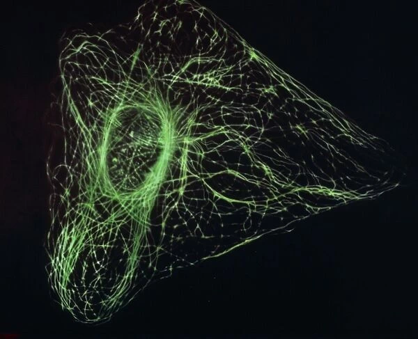
Cushion : Ultraviolet fluorescence micrograph animal cell
![]()

Home Decor from Science Photo Library
Ultraviolet fluorescence micrograph animal cell
Ultraviolet Fluorescence micrograph showing the microtubular network of an animal cell, made visible with fluorescent antibodies. Microtubules, together with microfilaments (not seen here), form a three-dimensional array known as the cytoskeleton of the cell. This fibrous network, only recently observed using ultraviolet light & fluorescent stains, is still poorly understood. Microtubules, more rigid than microfilaments, are thought to act as direction markers in the cell
Science Photo Library features Science and Medical images including photos and illustrations
Media ID 6401841
© FRANCIS LEROY, BIOCOSMOS/SCIENCE PHOTO LIBRARY
Cell Structure Cytology Cytoskeleton Microscopy Microtubule Micro Biology
Cushion
Refresh your home decor with a beautiful full photo 16"x16" (40x40cm) cushion, complete with cushion pad insert. Printed on both sides and made from 100% polyester with a zipper on the bottom back edge of the cushion cover. Care Instructions: Warm machine wash, do not bleach, do not tumble dry. Warm iron inside out. Do not dry clean.
Accessorise your space with decorative, soft cushions
Estimated Product Size is 40cm x 40cm (15.7" x 15.7")
These are individually made so all sizes are approximate
Artwork printed orientated as per the preview above, with landscape (horizontal) or portrait (vertical) orientation to match the source image.
EDITORS COMMENTS
This print from Science Photo Library showcases the intricate beauty of an animal cell's microtubular network. Through the use of fluorescent antibodies and ultraviolet fluorescence microscopy, this image reveals a three-dimensional array known as the cytoskeleton. The fibrous structure, which includes microtubules and microfilaments (not visible here), plays a crucial role in maintaining cell structure and facilitating cellular transport. The mesmerizing glow emitted by the fluorescent stains highlights the complexity of this biological marvel. While scientists have only recently been able to observe and study this network using ultraviolet light, its full functionality remains shrouded in mystery. Microtubules, with their rigid nature, are believed to act as directional markers within the cell. This photograph serves as a testament to the incredible advancements made in biology and microscopy. It reminds us that there is still much more to uncover about our own cells' inner workings. As we delve deeper into understanding these microscopic structures, we gain valuable insights into fundamental processes that sustain life itself. Science Photo Library has once again captured a moment of scientific wonder through this visually stunning print. It invites viewers to appreciate both the aesthetic appeal and scientific significance of this hidden world within us all.
MADE IN AUSTRALIA
Safe Shipping with 30 Day Money Back Guarantee
FREE PERSONALISATION*
We are proud to offer a range of customisation features including Personalised Captions, Color Filters and Picture Zoom Tools
SECURE PAYMENTS
We happily accept a wide range of payment options so you can pay for the things you need in the way that is most convenient for you
* Options may vary by product and licensing agreement. Zoomed Pictures can be adjusted in the Cart.


