Fine Art Print > Animals > Insects > Spiders > Yellow Sac
Fine Art Print : Artwork of section through the female bladder


![]()
Fine Art Prints from Science Photo Library
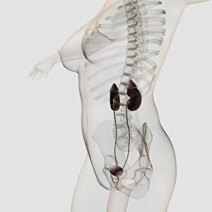
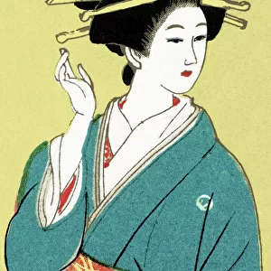
Artwork of section through the female bladder
Female bladder. Artwork showing a section through the female urinary system. The bladder is seen as a pink sac at centre. This is a hollow organ that holds urine. Two tubes (ureters) from upper image lead urine from the kidneys to the bladder. Yellow drops of urine are seen emerging from the trigone, the triangular space at the base of the bladder (lower centre). At the base of the bladder, the narrow urethra emerges, a tube that carries urine to the exterior opening. Just above the exterior opening, pink muscles of the urethral sphincter are seen. This muscle relaxes or contracts to control the flow of urine out of the bladder
Science Photo Library features Science and Medical images including photos and illustrations
Media ID 6451045
© JOHN BAVOSI/SCIENCE PHOTO LIBRARY
Bladder Sphincter Ureter Urethra Urinary Urinary System
20"x16" (+3" Border) Fine Art Print
Discover the intricacies of the human body with our Fine Art Prints from Media Storehouse. This captivating artwork, sourced from Science Photo Library, showcases a detailed section through the female urinary system. Witness the pink sac of the bladder at the center, holding the fluid that is essential for maintaining urinary health. Add this stunning piece to your collection and ignite curiosity in the wonders of anatomy.
20x16 image printed on 26x22 Fine Art Rag Paper with 3" (76mm) white border. Our Fine Art Prints are printed on 300gsm 100% acid free, PH neutral paper with archival properties. This printing method is used by museums and art collections to exhibit photographs and art reproductions.
Our fine art prints are high-quality prints made using a paper called Photo Rag. This 100% cotton rag fibre paper is known for its exceptional image sharpness, rich colors, and high level of detail, making it a popular choice for professional photographers and artists. Photo rag paper is our clear recommendation for a fine art paper print. If you can afford to spend more on a higher quality paper, then Photo Rag is our clear recommendation for a fine art paper print.
Estimated Image Size (if not cropped) is 40.6cm x 49.3cm (16" x 19.4")
Estimated Product Size is 55.9cm x 66cm (22" x 26")
These are individually made so all sizes are approximate
Artwork printed orientated as per the preview above, with portrait (vertical) orientation to match the source image.
FEATURES IN THESE COLLECTIONS
> Animals
> Insects
> Spiders
> Yellow Sac
EDITORS COMMENTS
This artwork showcases a detailed section through the female bladder, offering a fascinating glimpse into the intricate workings of the urinary system. At its core lies the pink sac-like structure, known as the bladder, which serves as a reservoir for urine. The upper part of this masterpiece reveals two slender tubes called ureters that transport urine from the kidneys to be stored in the bladder. A mesmerizing sight unfolds at the base of the bladder where drops of yellow-hued urine emerge from an area known as the trigone - a triangular space with significant physiological importance. Just below this remarkable phenomenon, we witness the emergence of another vital component: the narrow urethra. This tube acts as a conduit for carrying urine outwards towards its final destination. Drawing our attention further upwards, we encounter delicate pink muscles belonging to none other than the urethral sphincter. These muscles play a crucial role in regulating and controlling urination by either relaxing or contracting accordingly. Through this awe-inspiring artwork, Science Photo Library invites us on an intimate journey inside one aspect of human anatomy - specifically focusing on female physiology. It is truly remarkable how such intricacy exists within our bodies and serves as yet another testament to nature's extraordinary design.
MADE IN AUSTRALIA
Safe Shipping with 30 Day Money Back Guarantee
FREE PERSONALISATION*
We are proud to offer a range of customisation features including Personalised Captions, Color Filters and Picture Zoom Tools
SECURE PAYMENTS
We happily accept a wide range of payment options so you can pay for the things you need in the way that is most convenient for you
* Options may vary by product and licensing agreement. Zoomed Pictures can be adjusted in the Cart.