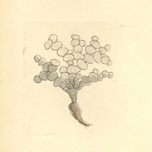
Fine Art Print > Animals > Mammals > Muridae > Water Mouse
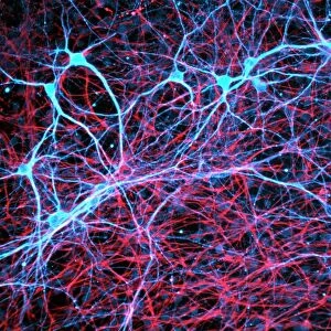
Fine Art Print : Brain fibres, DTI MRI scan C017 / 7036

![]()
Fine Art Prints from Science Photo Library
Brain fibres, DTI MRI scan C017 / 7036
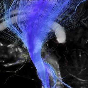
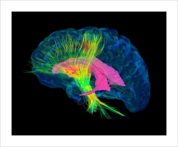
Brain fibres. 3D diffusion tensor imaging (DTI) magnetic resonance imaging (MRI) scan of a selection of nerve pathways (green/yellow) in the brain. The brain is seen from the side, with the front of the brain at left. The ventricles are pink and the brains cortical surface is blue. Diffusion tensor imaging measures the direction of water diffusion, which in the brain reveals the orientation of nerve fibres. The technique is also known as tractography, with the resulting image known as a tractogram
Science Photo Library features Science and Medical images including photos and illustrations
Media ID 9229819
© SHERBROOKE CONNECTIVITY IMAGING LAB/SCIENCE PHOTO LIBRARY
Brain Imaging Brain Scan Central Nervous System Cerebral Cerebrum Diffusion Tensor Imaging Dti Scan Fiber Fibers Fibre Fibres Imaging Technique Magnetic Resonance Imaging Mri Scan Mri Scanner Nerve Nerve Fibre Nerves Neural Pathway Neural Tract Paths Pathway Pathways Structural Tractogram Tractography Ventricle Ventricles White Matter Brain Neurological Neurology
20"x16" (+3" Border) Fine Art Print
Discover the intricacies of the human brain with our Fine Art Print from Media Storehouse, featuring the captivating image "Brain Fibres" by SHERBROOKE CONNECTIVITY IMAGING LAB/SCIENCE PHOTO LIBRARY. This stunning 3D diffusion tensor imaging (DTI) magnetic resonance imaging (MRI) scan reveals a mesmerizing network of nerve pathways in vibrant shades of green and yellow. Bring the wonders of neuroscience into your home or office with this beautiful, high-quality art print, a perfect conversation starter and an inspiring reminder of the complexities of the human mind.
20x16 image printed on 26x22 Fine Art Rag Paper with 3" (76mm) white border. Our Fine Art Prints are printed on 300gsm 100% acid free, PH neutral paper with archival properties. This printing method is used by museums and art collections to exhibit photographs and art reproductions.
Our fine art prints are high-quality prints made using a paper called Photo Rag. This 100% cotton rag fibre paper is known for its exceptional image sharpness, rich colors, and high level of detail, making it a popular choice for professional photographers and artists. Photo rag paper is our clear recommendation for a fine art paper print. If you can afford to spend more on a higher quality paper, then Photo Rag is our clear recommendation for a fine art paper print.
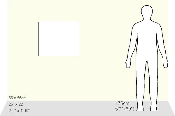
Estimated Image Size (if not cropped) is 50.8cm x 40.6cm (20" x 16")
Estimated Product Size is 66cm x 55.9cm (26" x 22")
These are individually made so all sizes are approximate
Artwork printed orientated as per the preview above, with landscape (horizontal) orientation to match the source image.
FEATURES IN THESE COLLECTIONS
> Animals
> Mammals
> Muridae
> Water Mouse
EDITORS COMMENTS
This print showcases the intricate network of brain fibres, captured through a cutting-edge imaging technique known as diffusion tensor imaging (DTI) magnetic resonance imaging (MRI). The image reveals a selection of nerve pathways in vibrant green and yellow hues against a striking black background. From this side view of the brain, with the front on the left, one can observe the pink ventricles and blue cortical surface. DTI measures water diffusion direction within the brain, providing valuable insights into the orientation of nerve fibres. This technique, also referred to as tractography, generates an image called a tractogram that beautifully illustrates these neural pathways. The photograph not only highlights the structural complexity and anatomical beauty of our brains but also serves as a reminder of their vital role in our overall health and well-being. It represents an amalgamation of biology, medicine, neurology, and anatomy – disciplines that rely on such advanced imaging techniques for research purposes. Captured by Sherbrooke Connectivity Imaging Lab from Science Photo Library's extensive collection, this mesmerizing print offers viewers a glimpse into the inner workings of our central nervous system. It is both scientifically informative and visually stunning—a testament to human ingenuity in unraveling nature's mysteries at microscopic levels.
MADE IN AUSTRALIA
Safe Shipping with 30 Day Money Back Guarantee
FREE PERSONALISATION*
We are proud to offer a range of customisation features including Personalised Captions, Color Filters and Picture Zoom Tools
SECURE PAYMENTS
We happily accept a wide range of payment options so you can pay for the things you need in the way that is most convenient for you
* Options may vary by product and licensing agreement. Zoomed Pictures can be adjusted in the Cart.