Fine Art Print > Arts > Artists > G > Thomas Gray
Fine Art Print : Purkinje nerve cells in the cerebellum


![]()
Fine Art Prints from Science Photo Library
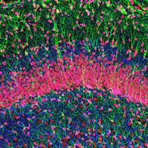
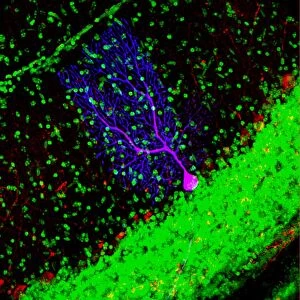
Purkinje nerve cells in the cerebellum
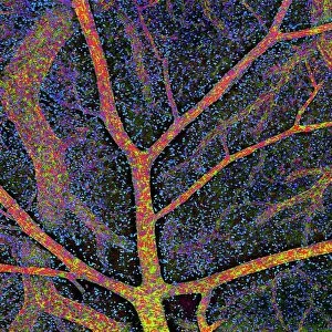
Purkinje cells in the cerebellum. Fluorescent light micrograph of Purkinje cells (green) in the cerebellum of the brain. Purkinje nerve cells have a flask-like body from which numerous highly branched dendrites extend. They are found in the grey matter (cortex) of the cerebellum, at the boundary between the granular layer (blue red) and the molecular layer (red green). The dendrites relay signals to the cell body, which passes them on through its single axon (green) in the granular layer. The cerebellum is a structure at the base of the brain that plays an important role in motor control, sensory perception and learning. Magnification: x380 when printed 10cm wide
Science Photo Library features Science and Medical images including photos and illustrations
Media ID 6448951
© THOMAS DEERINCK, NCMIR/SCIENCE PHOTO LIBRARY
Cerebellar Cerebellum Cortex Dendrite Dendrites Fluorescent Light Micrograph Granular Layer Gray Grey Matter Histological Histology Molecular Layer Nerve Cell Neuron Purkinje Cell Brain Light Microscope Nervous System Neurological Neurology
20"x20" (+3" Border) Fine Art Print
Discover the intricacies of the human body with our Fine Art Prints from Media Storehouse, featuring this captivating image of Purkinje nerve cells in the cerebellum. This mesmerizing fluorescent light micrograph, sourced from Science Photo Library, showcases the unique and complex structure of these essential brain cells. Bring the beauty of science into your home or office with this high-quality, museum-grade print. Each print is carefully crafted using archival inks and premium paper to ensure lasting brilliance and vibrancy. Immerse yourself in the wonders of the natural world and add a touch of intellectual elegance to your space with this stunning representation of Purkinje cells in the cerebellum.
20x20 image printed on 26x26 Fine Art Rag Paper with 3" (76mm) white border. Our Fine Art Prints are printed on 300gsm 100% acid free, PH neutral paper with archival properties. This printing method is used by museums and art collections to exhibit photographs and art reproductions.
Our fine art prints are high-quality prints made using a paper called Photo Rag. This 100% cotton rag fibre paper is known for its exceptional image sharpness, rich colors, and high level of detail, making it a popular choice for professional photographers and artists. Photo rag paper is our clear recommendation for a fine art paper print. If you can afford to spend more on a higher quality paper, then Photo Rag is our clear recommendation for a fine art paper print.
Estimated Image Size (if not cropped) is 50.8cm x 50.8cm (20" x 20")
Estimated Product Size is 66cm x 66cm (26" x 26")
These are individually made so all sizes are approximate
Artwork printed orientated as per the preview above, with landscape (horizontal) or portrait (vertical) orientation to match the source image.
FEATURES IN THESE COLLECTIONS
> Animals
> Mammals
> Muridae
> Blue-grey Mouse
> Arts
> Artists
> G
> Thomas Gray
> Science Photo Library
> Specialist Imaging
EDITORS COMMENTS
This print showcases the intricate beauty of Purkinje nerve cells in the cerebellum. The fluorescent light micrograph reveals these remarkable cells, with their distinct flask-like bodies and highly branched dendrites extending outwards. Positioned at the boundary between the granular layer and molecular layer in the grey matter of the cerebellum, these green-hued Purkinje cells play a crucial role in relaying signals. The image provides a glimpse into the complex inner workings of our brain's structure. With its single axon visible amidst the granular layer, this photograph highlights how these nerve cells transmit information from their dendrites to their cell body before passing it on further. The cerebellum itself holds immense significance as a vital component responsible for motor control, sensory perception, and learning. This microscopic view emphasizes its importance within our nervous system. With a magnification of x380 when printed 10cm wide, this print offers an up-close exploration into neurology and histology. It serves as a reminder of both our biological intricacies and the wonders that lie within us all.
MADE IN AUSTRALIA
Safe Shipping with 30 Day Money Back Guarantee
FREE PERSONALISATION*
We are proud to offer a range of customisation features including Personalised Captions, Color Filters and Picture Zoom Tools
SECURE PAYMENTS
We happily accept a wide range of payment options so you can pay for the things you need in the way that is most convenient for you
* Options may vary by product and licensing agreement. Zoomed Pictures can be adjusted in the Cart.