Framed Print : Underside of a slug, SEM


![]()
Framed Photos from Science Photo Library
Underside of a slug, SEM
Slug. Coloured scanning electron micrograph (SEM) of the underside of a slug (order Gastropoda). The underside of the slug is covered in microscopic hair-like projections known as cilia (yellow). Here the cilia are seen surrounded by mucus (slime, pink). Cilia beat in wave-like movements helping to propel the slug and disperse its slime
Science Photo Library features Science and Medical images including photos and illustrations
Media ID 6466157
© STEVE GSCHMEISSNER/SCIENCE PHOTO LIBRARY
Cilia Cilium False Colour Mucus Projection Projections Slime Slug Under Side Underneath False Coloured
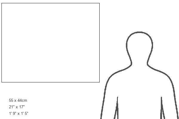
21.5"x17.5" (55x44cm) Premium Frame
Discover the intricacies of nature with Media Storehouse's Framed Prints featuring "Underside of a Slug, SEM" by Science Photo Library. This captivating image showcases the stunning detail of a slug's underside as captured through Scanning Electron Microscopy (SEM). Witness the mesmerizing microscopic hair-like projections, known as cilia, in vibrant yellow, covering the slug's surface. Bring the wonders of science into your home or office with this unique and thought-provoking addition to your décor.
Framed and mounted 17x12 print. Professionally handmade full timber moulded frames are finished off with framers tape and come with a hanging solution on the back. Outer dimensions are 21.5x17.5 inches (546x444mm). Quality timber frame frame moulding (20mm wide and 30mm deep) with frame colours in your choice of black, white, or raw oak and a choice of black or white card mounts. Frames have a perspex front providing a virtually unbreakable glass-like finish which is easily cleaned with a damp cloth.
Contemporary Framed and Mounted Prints - Professionally Made and Ready to Hang
Estimated Image Size (if not cropped) is 41.8cm x 41.8cm (16.5" x 16.5")
Estimated Product Size is 54.6cm x 44.4cm (21.5" x 17.5")
These are individually made so all sizes are approximate
Artwork printed orientated as per the preview above, with landscape (horizontal) or portrait (vertical) orientation to match the source image.
EDITORS COMMENTS
This print showcases the mesmerizing world beneath a slug's slimy exterior. Taken with a scanning electron microscope, the image reveals intricate details of the slug's underside that are otherwise invisible to the naked eye. The vibrant colors bring to life the fascinating biology of this creature. The focal point of this image is undoubtedly the countless cilia covering the slug's body. These hair-like projections, depicted in striking yellow hues, play a crucial role in its locomotion and slime dispersal. As we observe closely, we can witness these cilia beating in synchronized wave-like movements, propelling the slug forward and aiding its movement through various terrains. Adding an extra layer of intrigue to this composition is the presence of mucus or slime surrounding each cilium. Delicately colored in pink tones, it serves as both protection and lubrication for our gastropod friend. This photograph not only highlights nature's remarkable diversity but also emphasizes how even seemingly insignificant organisms possess extraordinary adaptations for survival. It invites us into an unseen realm where microscopic wonders await exploration. With its blend of artistry and scientific precision, this print from Science Photo Library offers a glimpse into one small corner of our vast natural world—an invitation to marvel at Earth's incredible biodiversity and appreciate every living being’s unique place within it.
MADE IN AUSTRALIA
Safe Shipping with 30 Day Money Back Guarantee
FREE PERSONALISATION*
We are proud to offer a range of customisation features including Personalised Captions, Color Filters and Picture Zoom Tools
SECURE PAYMENTS
We happily accept a wide range of payment options so you can pay for the things you need in the way that is most convenient for you
* Options may vary by product and licensing agreement. Zoomed Pictures can be adjusted in the Cart.