Fraud Blocker Skip to main content

Cell Cycle Collection

The cell cycle is a fascinating process that governs the growth and development of living organisms

Background imageCell Cycle Collection: Embryo development 24-36 hours after fertilization

Embryo development 24-36 hours after fertilization

Background imageCell Cycle Collection: Conceptual image of RNA virus replication

Conceptual image of RNA virus replication

Background imageCell Cycle Collection: Zygote development 24-36 hours after fertilization

Zygote development 24-36 hours after fertilization

Background imageCell Cycle Collection: Conceptual image of mitosis

Conceptual image of mitosis. Mitosis is the process in the cell cycle by which a cell duplicates into two genetically identical sets of chromosomes

Background imageCell Cycle Collection: Conceptual image of cell nucleus

Conceptual image of cell nucleus. The cell nucleus acts like the brain of the cell. It helps control eating, movement, and reproduction

Background imageCell Cycle Collection: Microscopic view of cell division

Microscopic view of cell division

Background imageCell Cycle Collection: Tumour suppressor protein and DNA C017 / 3647

Tumour suppressor protein and DNA C017 / 3647
Tumour suppressor protein and DNA. Computer artwork showing a molecule of the tumour suppressor protein p53 (blue and pink) bound to a molecule of DNA (deoxyribonucleic acid, yellow and orange)

Background imageCell Cycle Collection: Sirtuin enzyme and p53, artwork C017 / 3659

Sirtuin enzyme and p53, artwork C017 / 3659
Sirtuin enzyme and p53. Computer artwork of a sirtuin (Sir2) enzyme (pink) bound to a p53 peptide (orange). Sir2 enzymes form a unique class of NAD(+)

Background imageCell Cycle Collection: Tumour suppressor protein and DNA C017 / 3644

Tumour suppressor protein and DNA C017 / 3644
Tumour suppressor protein and DNA. Computer artwork showing a molecule of the tumour suppressor protein p53 (blue and pink) bound to a molecule of DNA (deoxyribonucleic acid, yellow and orange)

Background imageCell Cycle Collection: Sirtuin enzyme and p53, artwork C017 / 3658

Sirtuin enzyme and p53, artwork C017 / 3658
Sirtuin enzyme and p53. Computer artwork of a sirtuin (Sir2) enzyme (pink) bound to a p53 peptide (orange). Sir2 enzymes form a unique class of NAD(+)

Background imageCell Cycle Collection: Tumour suppressor protein and DNA C017 / 3645

Tumour suppressor protein and DNA C017 / 3645
Tumour suppressor protein and DNA. Computer artwork showing a molecule of the tumour suppressor protein p53 (blue and pink) bound to a molecule of DNA (deoxyribonucleic acid, yellow and orange)

Background imageCell Cycle Collection: Sirtuin enzyme and p53, artwork C017 / 3660

Sirtuin enzyme and p53, artwork C017 / 3660
Sirtuin enzyme and p53. Computer artwork of a sirtuin (Sir2) enzyme (blue) bound to a p53 peptide (pink). Sir2 enzymes form a unique class of NAD(+)

Background imageCell Cycle Collection: Tumour suppressor protein with DNA F006 / 9729

Tumour suppressor protein with DNA F006 / 9729
Tumour suppressor protein. Molecular model of the tumour suppressor protein p53 (beige) bound to a molecule of DNA (deoxyribonucleic acid, red and blue)

Background imageCell Cycle Collection: Tumour suppressor protein with DNA F006 / 9564

Tumour suppressor protein with DNA F006 / 9564
Tumour suppressor protein. Molecular model of the tumour suppressor protein p53 (beige) bound to a molecule of DNA (deoxyribonucleic acid, red and blue)

Background imageCell Cycle Collection: Tumour suppressor protein with DNA F006 / 9523

Tumour suppressor protein with DNA F006 / 9523
Tumour suppressor protein. Molecular model of the tumour suppressor protein p53 (beige) bound to a molecule of DNA (deoxyribonucleic acid, red and blue)

Background imageCell Cycle Collection: Tumour suppressor protein molecular model F006 / 9450

Tumour suppressor protein molecular model F006 / 9450
Tumour suppressor protein, molecular model. P53 prevents the proliferation of cells with damaged DNA. Its production occurs in response to radiation and chemicals that damage the structure of DNA

Background imageCell Cycle Collection: Oxoguanine glycosylase complex F006 / 9318

Oxoguanine glycosylase complex F006 / 9318
Oxoguanine glycosylase complex. Computer model showing an 8-Oxoguanine glycosylase (OGG1) molecule (beige) bound to a section of DNA (deoxyribonucleic acid, red and blue)

Background imageCell Cycle Collection: Oxoguanine glycosylase complex F006 / 9307

Oxoguanine glycosylase complex F006 / 9307
Oxoguanine glycosylase complex. Computer model showing an 8-Oxoguanine glycosylase (OGG1) molecule (beige) bound to a section of DNA (deoxyribonucleic acid, red and blue)

Background imageCell Cycle Collection: Dividing cell, TEM

Dividing cell, TEM
Dividing cell. Transmission electron micrograph (TEM) of a section through a cell dividing through the process of mitosis

Background imageCell Cycle Collection: Cyclin-dependent kinase inhibitor complex

Cyclin-dependent kinase inhibitor complex. Molecular model of the inhibitor protein P27Kip1 bound to a cyclin-A-cyclin dependent kinase 2 complex

Background imageCell Cycle Collection: Meiosis, illustration C018 / 0803

Meiosis, illustration C018 / 0803
Meiosis. Illustration of a cell during metaphase (I) of meiosis (gamete formation). During meiosis four daughter nuclei are formed from one parent nucleus after two stages of nuclear division

Background imageCell Cycle Collection: Yeast cells, illustration C018 / 0750

Yeast cells, illustration C018 / 0750
Yeast cells. Illustration of cells of brewer s, or baker s, yeast (Saccharomyces cerevisiae). This fungus consists of single vegetative cells

Background imageCell Cycle Collection: Yeast cells, illustration C018 / 0752

Yeast cells, illustration C018 / 0752
Yeast cells. Illustration of cells of brewer s, or baker s, yeast (Saccharomyces cerevisiae). This fungus consists of single vegetative cells. S

Background imageCell Cycle Collection: Tumour suppressor protein and DNA C017 / 3643

Tumour suppressor protein and DNA C017 / 3643
Tumour suppressor protein and DNA. Computer artwork showing a molecule of the tumour suppressor protein p53 (blue and pink) bound to a molecule of DNA (deoxyribonucleic acid, yellow and orange)

Background imageCell Cycle Collection: Dividing cancer cell, SEM C014 / 0362

Dividing cancer cell, SEM C014 / 0362
Dividing cancer cell. Coloured scanning electron micrograph (SEM) of a colorectal cancer cell undergoing mitosis (nuclear division) and splitting into two daughter cells (left and right)

Background imageCell Cycle Collection: Mouth cancer cell dividing, SEM

Mouth cancer cell dividing, SEM
Mouth cancer dividing. Coloured scanning electron micrograph (SEM) of a squamous cell carcinoma (cancer) cell from a human mouth undergoing mitosis (nuclear division)

Background imageCell Cycle Collection: Dividing liver cancer cell, SEM

Dividing liver cancer cell, SEM
Dividing liver cancer cell. Coloured scanning electron micrograph (SEM) of a hepatocellular carcinoma (HCC) cell undergoing mitosis (nuclear division)

Background imageCell Cycle Collection: Dividing cancer cell, SEM C014 / 0361

Dividing cancer cell, SEM C014 / 0361
Dividing cancer cell. Coloured scanning electron micrograph (SEM) of a colorectal cancer cell undergoing mitosis (nuclear division) and splitting into two daughter cells (left and right)

Background imageCell Cycle Collection: Dividing brain cancer cells, SEM C014 / 0354

Dividing brain cancer cells, SEM C014 / 0354
Dividing brain cancer cells. Coloured scanning electron micrograph (SEM) of a cancerous astrocyte brain cell that has just undergone cytokinesis (cell division)

Background imageCell Cycle Collection: Dividing bone cancer cell, SEM

Dividing bone cancer cell, SEM
SEM Dividing Osteosarcoma cells Osteosarcoma is an aggressive malignant neoplasm arising from primitive transformed cells of mesenchymal origin (and thus a sarcoma)

Background imageCell Cycle Collection: Tumour suppressor protein molecular model C016 / 2065

Tumour suppressor protein molecular model C016 / 2065
Tumour suppressor protein. Molecular model of the tumour suppressor protein p53 (left and right) bound to a molecule of DNA (deoxyribonucleic acid, down centre) at the p53 response element

Background imageCell Cycle Collection: Importin protein complexed with enzyme C015 / 5149

Importin protein complexed with enzyme C015 / 5149
Importin protein complexed with enzyme, molecular model. Human importin13 (purple) complexed with SUMO-conjugating enzyme Ubc9 (red)

Background imageCell Cycle Collection: Importin protein complexed with enzyme C015 / 5148

Importin protein complexed with enzyme C015 / 5148
Importin protein complexed with enzyme, molecular model. Human importin13 (green) complexed with SUMO-conjugating enzyme Ubc9 (pink)

Background imageCell Cycle Collection: DNA and tumour suppressor complex C014 / 0014

DNA and tumour suppressor complex C014 / 0014
DNA and tumour suppressor complex. Molecular model showing a molecule of the p53 tumour suppressor protein (green, centre) bound to a damaged DNA (deoxyribonucleic acid) strand

Background imageCell Cycle Collection: Oxoguanine glycosylase complex C013 / 8886

Oxoguanine glycosylase complex C013 / 8886
Oxoguanine glycosylase complex. Computer model showing a molecule of human aG DNA repair glycosylase (right) bound to an DNA molecule (left)

Background imageCell Cycle Collection: Oxoguanine glycosylase complex C013 / 8884

Oxoguanine glycosylase complex C013 / 8884
Oxoguanine glycosylase complex. Computer model showing an 8-Oxoguanine glycosylase (OGG1) molecule (green) bound to a section of DNA (deoxyribonucleic acid, pink and blue)

Background imageCell Cycle Collection: Tumour suppressor protein molecular model C013 / 7914

Tumour suppressor protein molecular model C013 / 7914
Tumour suppressor protein. Molecular model of the tumour suppressor protein p53 (blue, green and orange) bound to a molecule of DNA (deoxyribonucleic acid, yellow and pink)

Background imageCell Cycle Collection: Cell division, SEM

Cell division, SEM
Cell division. Coloured scanning electron micrograph (SEM) of two animal cells during cytokinesis (cell division). Cytokinesis occurs after nuclear division (mitosis)

Background imageCell Cycle Collection: Cyclin-dependent kinase 2 enzyme

Cyclin-dependent kinase 2 enzyme, molecular model. This enzyme is found in cells, where it is involved in regulating the cell cycle, the cycle of cell division and cell growth


All Professionally Made to Order for Quick Shipping

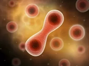
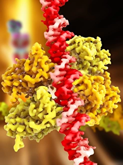
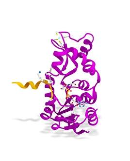
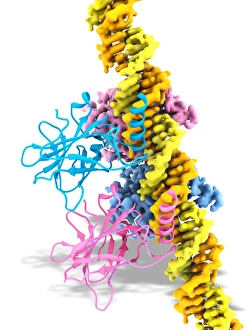
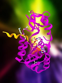
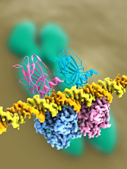
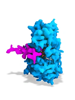
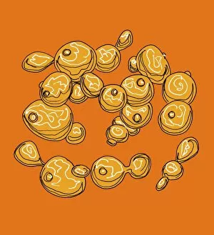
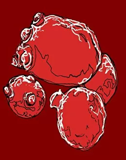
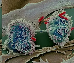
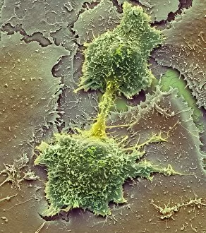
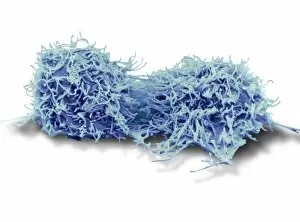
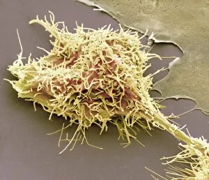
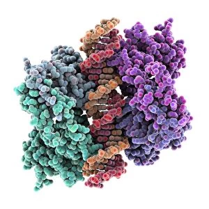
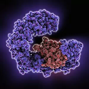
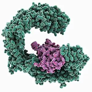
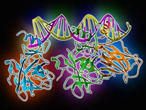
The cell cycle is a fascinating process that governs the growth and development of living organisms. From the early stages of embryo development, where cells divide rapidly 24-36 hours after fertilization, to the intricate replication of RNA viruses, this cycle showcases the complexity and beauty of life. Imagine peering through a scanning electron microscope (SEM) and witnessing cell division in action. The microscopic view reveals an awe-inspiring sight as cells split into two, each carrying genetic information vital for future growth. In parallel, zygote development unfolds within the same time frame after fertilization. This crucial stage lays the foundation for an organism's entire existence by merging genetic material from both parents. Conceptual images provide us with glimpses into other aspects of the cell cycle. One captures mitosis - a breathtaking dance where chromosomes align and separate flawlessly to ensure accurate distribution during cellular reproduction. Another focuses on the intricate structure of a cell nucleus, housing DNA that holds our unique traits and characteristics. But it's not just about growth; maintaining balance is equally important. Tumour suppressor proteins play a critical role in preventing uncontrolled cell division by monitoring DNA integrity closely. Artwork depicting their interaction with DNA highlights their significance in safeguarding against harmful mutations. Sirtuin enzymes also come into play when it comes to regulating cellular processes like aging and stress response. Their partnership with p53 protein helps maintain genomic stability, ensuring healthy functioning throughout our lives. As we delve deeper into understanding these mechanisms at work within our bodies, we gain insights into how life thrives amidst constant change. The captivating visuals provided by scientific research fuel our curiosity while reminding us of nature's remarkable intricacies. So next time you ponder over life's mysteries or marvel at its diversity, remember that behind every concept image or microscopic view lies another piece unraveling the wonders hidden within the incredible journey called the cell cycle.