Fraud Blocker Skip to main content

Pathology Collection (#11)

"Exploring the Intricate World of Pathology: Unveiling the Mysteries Within" Delving into the realm of pathology, we encounter a fascinating array of scientific wonders

Background imagePathology Collection: Rust fungus, SEM

Rust fungus, SEM
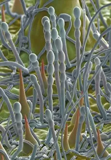
Rust fungus. Coloured scanning electron micrograph (SEM) of spores of the mint rust fungus (Puccinia menthae, orange) on the surface of a peppermint leaf (Mentha piperita)

Background imagePathology Collection: Powdery mildew, SEM

Powdery mildew, SEM
Powdery mildew. Coloured scanning electron micrograph (SEM) of the parasitic powdery mildew Erysiphe sp. on the stalk of a plant

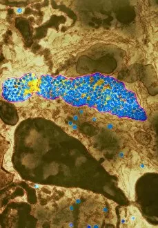
Background imagePathology Collection: HIV particles in infected cell, TEM

HIV particles in infected cell, TEM
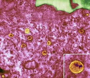
HIV particles. Coloured transmission electron micrograph (TEM) of human immunodeficiency virus (HIV) particles (orange) in a host cell. HIV causes the disease AIDS

Background imagePathology Collection: Freezing human brain slices

Freezing human brain slices
Human brain sections being frozen. A technician pours liquid nitrogen onto human brain tissue. The brains have been donated for medical research at the Harvard Brain and Tissue Resource Centre, USA

Background imagePathology Collection: Technician dissecting a human brain

Technician dissecting a human brain. A technician slices a human brain into 1.5 centimetre thick sections. It has been donated for medical research at the Harvard Brain and Tissue Resource Centre

Background imagePathology Collection: Recording body slice for Visible Human project

Recording body slice for Visible Human project
Morbid anatomy. A scientist prepares to photograph a transverse section of a human head, using cameras at the top of the picture

Background imagePathology Collection: Crystals in seminal fluid

Crystals in seminal fluid. Polarised light micrograph of a sample of abnormal seminal fluid. The sample contains crystals, which may indicate a problem with the prostate

Background imagePathology Collection: Human brain dissected in half

Human brain dissected in half. A human brain dissected into right and left hemispheres. It has been donated for medical research at the Harvard Brain and Tissue Resource Centre, USA

Background imagePathology Collection: Bacterial infection of nail, SEM

Bacterial infection of nail, SEM
Bacterial infection of a nail. Coloured scanning electron micrograph (SEM) of a nail with a bacterial infection (paronychia)

Background imagePathology Collection: Bleeding in emphysema

Bleeding in emphysema
Internal bleeding in lung tissue due to pulmonary emphysema, light micrograph. The bleeding is seen as numerous red blood cells (red)

Background imagePathology Collection: Studying the brain, conceptual image

Studying the brain, conceptual image

Background imagePathology Collection: Brains in petri dishes, conceptual image

Brains in petri dishes, conceptual image
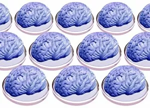
Brains in petri dishes, conceptual artwork

Background imagePathology Collection: Neutrophil cell and bacteria, SEM

Neutrophil cell and bacteria, SEM
Neutrophil cell and bacteria. Coloured scanning electron micrograph (SEM) of bacteria (rod-shaped) and part of a neutrophil cell (lower left)

Background imagePathology Collection: Macrophage cell engulfing bacteria, TEM

Macrophage cell engulfing bacteria, TEM
Macrophage cell engulfing bacteria. Coloured transmission electron micrograph (TEM) of bacteria (centre, rod-shaped) inside a macrophage cell

Background imagePathology Collection: Back deformities

Back deformities, historical anatomical artwork. These patients all have spinal deformities leading to abnormal curvatures of the back

Background imagePathology Collection: Aortic aneurysm

Aortic aneurysm. Artwork from Jean Cruveilhiers medical textbook Anatomie pathalogique du corps humain, published 1835. The two large illustrations are views of an aortic aneurysm (brown, centre)

Background imagePathology Collection: Adenovirus, TEM

Adenovirus, TEM

Background imagePathology Collection: AIDS in the UK

AIDS in the UK, conceptual image. Computer artwork of red ribbons overlain on a map of the UK. These ribbons are worn to show support for the cause of AIDS prevention

Background imagePathology Collection: People with AIDS

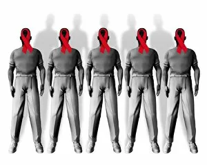
People with AIDS, conceptual image. Computer artwork of red ribbons overlaid on the faces of people with AIDS. These ribbons are worn to show support for the cause of AIDS prevention

Background imagePathology Collection: Dengue fever virus particles, TEM

Dengue fever virus particles, TEM

Background imagePathology Collection: Dengue fever virus replication, TEM

Dengue fever virus replication, TEM

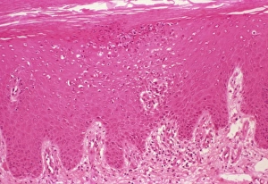
Background imagePathology Collection: Psoriasis, light micrograph

Psoriasis, light micrograph
Psoriasis. Light micrograph of a section through a psoriasis pustule. Psoriasis is a chronic skin disease causing inflammation and scaly lesions

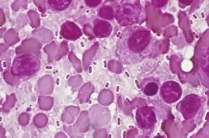
Background imagePathology Collection: Bacterial pneumonia infection

Bacterial pneumonia infection. Light micrograph of pneumonia bacteria (Klebsiella pneumoniae, rod- shaped, pink) among cells in a sample of pus from a pneumonia patient. K

Background imagePathology Collection: World AIDS epidemic

World AIDS epidemic, conceptual image. Computer artwork of red ribbons overlain on a map of the world. These ribbons are worn to show support for the cause of AIDS prevention

Background imagePathology Collection: Edme Vulpian, French neurologist

Edme Vulpian, French neurologist
Edme Felix Alfred Vulpian (1826-1887), French neurologist. Vulpian was born and educated in Paris. His doctoral thesis (1853) was on the origin of cranial nerves III to X

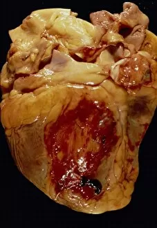
Background imagePathology Collection: Ruptured heart

Ruptured heart. Gross specimen of a heart that has ruptured in the parietal region (dark blood clots, lower centre, and red region, centre)

Background imagePathology Collection: Typhoid nodules, light micrograph

Typhoid nodules, light micrograph
Typhoid nodules, coloured light micrograph. Section through a lymph node of a patient with typhoid, showing macrophages (large cells, pink) forming typhoid nodules

Background imagePathology Collection: Liver tuberculosis, light micrograph

Liver tuberculosis, light micrograph
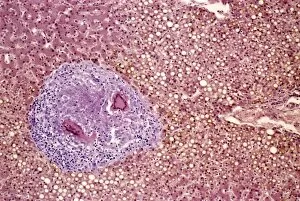
Liver tuberculosis. Coloured light micrograph of a section through the liver of a patient with miliary tuberculosis (TB). A tubercle, a nodular lesion of infected dead tissue, is seen at left

Background imagePathology Collection: Miliary tuberculosis, light micrograph

Miliary tuberculosis, light micrograph
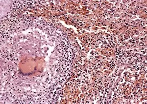
Miliary tuberculosis. Light micrograph of a section through a single miliary tubercle (left) in a patients lung. Tubercles are nodular lesions of infected dead tissue that arise from tuberculosis (TB)

Background imagePathology Collection: Demyelinated nerve, TEM

Demyelinated nerve, TEM
Demyelinated nerve. Coloured transmission electron micrograph (TEM) of a section through an axon (a structure that transmits nerve impulses to other nerve cells) that has lost its myelin sheath

Background imagePathology Collection: Demyelinated nerve in multiple sclerosis

Demyelinated nerve in multiple sclerosis. Coloured transmission electron micrograph (TEM) of a section through an axon (a structure that transmits nerve impulses) that has lost its myelin sheath

Background imagePathology Collection: Multiple sclerosis, SEM

Multiple sclerosis, SEM
Multiple sclerosis. Coloured scanning electron micrograph (SEM) of microglial cells (round) ingesting oligodendrocytes (branched). This is the process thought to occur in multiple sclerosis (MS)

Background imagePathology Collection: Systemic lupus erythematosus

Systemic lupus erythematosus. Light micrograph of a section through a glomerulus (blood filtration structure) from kidney tissue in a case of SLE

Background imagePathology Collection: Felix Guyon, French urologist

Felix Guyon, French urologist

Background imagePathology Collection: Giovanni Battista Morgagni, anatomist

Giovanni Battista Morgagni, anatomist

Background imagePathology Collection: Lung fibrosis, CT scan

Lung fibrosis, CT scan
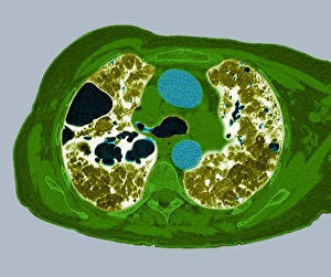
Lung fibrosis. Image 2 of 3. Coloured computed tomography (CT) scan through the chest of a patient with lung (pulmonary) fibrosis

Background imagePathology Collection: Creutzfeldt-Jakob diseased brain

Creutzfeldt-Jakob diseased brain
Creutzfeldt-Jakob disease (CJD). Computer artwork based on a light micrograph of a section through a human brain exhibiting CJD

Background imagePathology Collection: Crohns disease, X-ray

Crohns disease, X-ray

Background imagePathology Collection: Skin inflammation, light micrograph

Skin inflammation, light micrograph
Skin inflammation. Light micrograph of a section through skin showing inflamed capillaries (lower left and upper right) caused by capillaritis, also known as pigmented purpurea


All Professionally Made to Order for Quick Shipping

"Exploring the Intricate World of Pathology: Unveiling the Mysteries Within" Delving into the realm of pathology, we encounter a fascinating array of scientific wonders. From a set of glass eyeballs that have witnessed countless medical marvels to human brain microscope slides revealing intricate neural networks, each specimen holds secrets waiting to be unraveled. Intriguingly, a tobacco hornworm intertwined with its tobacco plant counterpart reminds us of the intricate relationship between organisms and their environment. Similarly, an Alzheimer's brain reveals the devastating effects this disease has on our most precious organ. As we journey through history, we encounter Louis Pasteur, the renowned French microbiologist whose groundbreaking discoveries paved the way for modern medicine. His contributions continue to inspire generations in their pursuit of understanding diseases at their core. Peering through microscopes, we witness captivating views such as that of a microscopic respiratory syncytial virus invading its host or Dohle bodies within blood cells – tiny anomalies holding vital diagnostic clues. The microscopic view of chlamydia serves as a reminder that even seemingly inconspicuous pathogens can wreak havoc on our health. Examining acute promyelocytic leukemia under magnification brings awareness to the battle fought by patients and healthcare professionals alike against this aggressive form of cancer. Meanwhile, glimpsing at sleeping sickness parasites highlights how these minuscule invaders disrupt lives in regions plagued by poverty and limited resources. The sight of liver cirrhosis evokes contemplation about lifestyle choices and their consequences on one's vital organs. In contrast, light micrographs showcasing liver tissue cirrhosis provide insight into cellular changes occurring within this complex disease process. Pathology is not merely confined to textbooks; it is an ever-evolving field where scientists tirelessly strive to decipher nature's enigmatic puzzles. Each slide represents more than just pixels; they hold stories waiting to be told – tales that shed light on diseases afflicting humanity and offer hope for better treatments.