Fraud Blocker Skip to main content

Plant Anatomy Collection

"Exploring the Intricate World of Plant Anatomy: A Fascinating Journey through Micrographs and Artwork" Delve into the hidden wonders with these captivating images

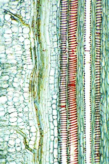
Background imagePlant Anatomy Collection: Dicotyledon plant stem, light micrograph

Dicotyledon plant stem, light micrograph
Dicotyledon plant stem. Light micrograph of a longitudinal radial section through a typical dicotyledon stem. The section has passed through a vascular bundle and other stem tissue

Background imagePlant Anatomy Collection: Castor oil stem, light micrograph

Castor oil stem, light micrograph
Castor oil stem. Light micrograph of a longitudinal section through the stem of a castor oil (Ricinus communis) plant. At right are large and small parenchyma cells (blue)

Background imagePlant Anatomy Collection: Maize root, light micrograph

Maize root, light micrograph
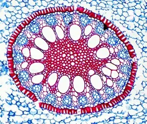
Maize root. Light micrograph of a section through the root of a maize plant (Zea mays) showing a vascular cylinder (centre)

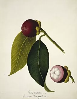
Background imagePlant Anatomy Collection: Mangosteen (Garcinia mangostana) C016 / 5659

Mangosteen (Garcinia mangostana) C016 / 5659
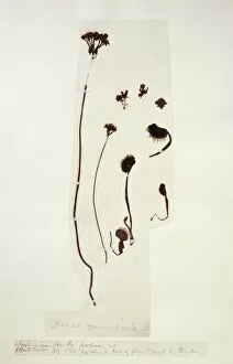
Mangosteen (Garcinia mangostana). Plate 35 by Margaret Bushby Lascelles Cockburn (1829-1928). Held at the Natural History Museum, London, UK

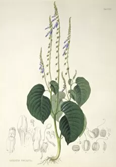
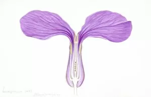
Background imagePlant Anatomy Collection: Rhynchoglossum obliquum, artwork C016 / 5646

Rhynchoglossum obliquum, artwork C016 / 5646
Rhynchoglossum obliquum. Engraving by Franz Bauer (from a drawing by Ferdinand Bauer) published in Plantae javanicae raroriores (1838)

Background imagePlant Anatomy Collection: English oak leaf pores, SEM

English oak leaf pores, SEM
English oak leaf pores. Coloured scanning electron micrograph (SEM) of stomata (round) on the underside of a leaf from an English oak (Quercus robur) tree

Background imagePlant Anatomy Collection: Cotton plant leaf, light micrograph

Cotton plant leaf, light micrograph
Cotton plant leaf. Light micrograph of a transverse section through a leaf of the cotton plant (Gossypium hirsutum). Magnification: x45 when printed at 10 centimetres wide

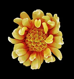
Background imagePlant Anatomy Collection: Buttercup flower, SEM

Buttercup flower, SEM
Buttercup flower (Ranunculus sp.), coloured scanning electron micrograph (SEM). The pistil (centre) is surrounded by the stamens (yellow)

Background imagePlant Anatomy Collection: Tomato leaf, light micrograph

Tomato leaf, light micrograph
Tomato leaf. Light micrograph of a transverse section through the midrib of a tomato (Lycopersicum esculentum) leaf. The upper and lower epidermis on the surfaces of the leaf are blue

Background imagePlant Anatomy Collection: Pollination, SEM

Pollination, SEM
Pollination. Coloured scanning electron micrograph (SEM) of part of a flowers pistil with pollen grains attached. The pistil is the female reproductive part of a flower

Background imagePlant Anatomy Collection: French lavender leaf pore, SEM

French lavender leaf pore, SEM
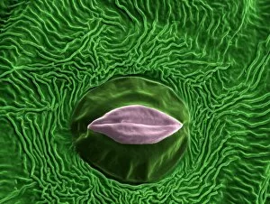
French lavender leaf pore. Coloured scanning electron micrograph (SEM) of an open stoma (centre, black). Stomata are pores that open and close in order to regulate gas exchange in a plant

Background imagePlant Anatomy Collection: Hand painted botanical print

Hand painted botanical print
Hand painted botanical study of a Costus villosissimus flower and leaf anatomy from Fragmenta Botanica by Nikolaus Joseph Freiherr von Jacquin or Baron Nikolaus von Jacquin (printed in Vienna in 1809)

Background imagePlant Anatomy Collection: Chloroplast, SEM

Chloroplast, SEM
Chloroplast. Coloured scanning electron micrograph (SEM) of a section through a plant cell, showing a fractured chloroplast (green)

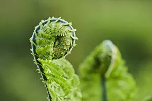
Background imagePlant Anatomy Collection: Unfurling of a Worm Fern

Unfurling of a Worm Fern
Close-up detail of the curled end of a male fern or worm fern (Dryopteris filix-mas); Bavaria, Germany

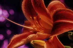
Background imagePlant Anatomy Collection: Daylilies, Try It, New York Botanical Garden, Bronx, New York, USA

Daylilies, Try It, New York Botanical Garden, Bronx, New York, USA
Daylilies (Hemerocallis) Try It, New York Botanical Garden. Digitized from film; Bronx, New York, United States of America

Background imagePlant Anatomy Collection: Common hepatica blossoms

Common hepatica blossoms
Common hepatica (Anemone hepatica) flowering in a forest; Upper Palatinate, Bavaria, Germany

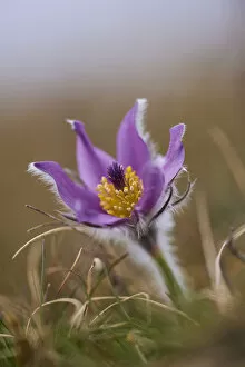
Background imagePlant Anatomy Collection: European pasqueflower in full bloom

European pasqueflower in full bloom
European pasqueflower (Pulsatilla vulgaris) in full bloom; Bavaria, Germany

Background imagePlant Anatomy Collection: Siberian squill blossoms

Siberian squill blossoms
Siberian squill or wood squill (Scilla siberica) blossoms; Bavaria, Germany

Background imagePlant Anatomy Collection: Daylilies, Satin Silk, New York Botanical Garden, Bronx, New York, USA

Daylilies, Satin Silk, New York Botanical Garden, Bronx, New York, USA
Daylilies (Hemerocallis) Satin Silk, New York Botanical Garden. Digitized from film; Bronx, New York, United States of America

Background imagePlant Anatomy Collection: White Easter Lilies, Snow Queen, New York Botanical Garden, Bronx, New York, USA

White Easter Lilies, Snow Queen, New York Botanical Garden, Bronx, New York, USA
White Easter Lilies (Lilium) Snow Queen Longiflorrum Liliaceae, New York Botanical Garden. Digitized from film; Bronx, New York, United States of America

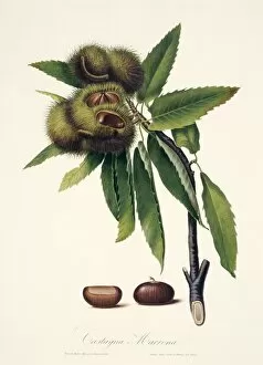
Background imagePlant Anatomy Collection: Sweet chestnut, 19th century illustration

Sweet chestnut, 19th century illustration
Illustration of sweet chestnut (Castanea sativa) fruits and nuts. Plate from Pomona Italiana, Vol 2 (1839) by Giorgio Galisio

Background imagePlant Anatomy Collection: Potato Leaf Stomata (SEM)

Potato Leaf Stomata (SEM)
Potato Stomata. Scanning electron micrographs (SEM) of open stomata on a potato leaf (Solanum tuberosum). Stomata are pores that open and close in order to regulate gas exchange in a plant

Background imagePlant Anatomy Collection: Iris root, light micrograph

Iris root, light micrograph
Iris root. Light micrograph of a section through the root of an Iris plant (Iris germanica) showing a vascular cylinder. The cylinder is comprised of a central cluster of parenchyma cells (red)

Background imagePlant Anatomy Collection: Plants and photosynthesis C014 / 1252

Plants and photosynthesis C014 / 1252
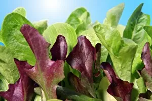
Plants and photosynthesis. Sunlight (upper left) shining on leafy plants, representing photosynthesis. This is the process by which plants harness the energy in sunlight

Background imagePlant Anatomy Collection: Algae cell wall, SEM

Algae cell wall, SEM
Algae cell wall. Coloured scanning electron micrograph (SEM) of cellulose microfibrils (strands) in a Chaetomorpha sp. algal cell wall

Background imagePlant Anatomy Collection: Plant stoma, ESEM

Plant stoma, ESEM
Plant stoma, coloured environmental scanning electron micrograph (ESEM). A stoma is a pore that regulates the exchange of gases and water vapour into and out of the plant

Background imagePlant Anatomy Collection: Venus flytrap (Dionaea muscipula) C016 / 5564

Venus flytrap (Dionaea muscipula) C016 / 5564
Venus flytrap (Dionaea muscipula) specimen collected by William Bartram (1739-1823)

Background imagePlant Anatomy Collection: ginger (Zingiber officinale), artwork C016 / 6130

ginger (Zingiber officinale), artwork C016 / 6130
Ginger (Zingiber officinale). Plate 909 from the John Reeves Collection of Botanical Drawings from Canton, China. Held in the Botany Library at the Natural History Museum, London, UK

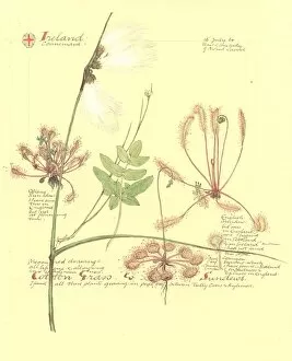
Background imagePlant Anatomy Collection: Eriophorum sp. and Drosera sp. artwork C016 / 5940

Eriophorum sp. and Drosera sp. artwork C016 / 5940
cotton grass (Eriophorum sp.) and sundew (Drosera sp.) plants. Drawing from Watercolour and pencil drawings of birds, mammals and boats of the Shetland Islands

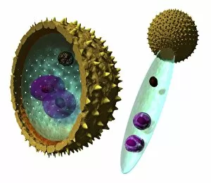
Background imagePlant Anatomy Collection: Pollen cell structures, artwork

Pollen cell structures, artwork
Pollen cell structures. Pollen grains are the male reproductive structures produced by flowering plants. They have a double wall (intine and exine)

Background imagePlant Anatomy Collection: Fern (Polypodium vulgare), artwork C016 / 6010

Fern (Polypodium vulgare), artwork C016 / 6010
Fern (Polypodium vulgare). Page 2 from The Ferns of Sidmouth (1862) by Peter Orlando Hutchinson. Held in the Botany Library at the Natural History Museum, London, UK

Background imagePlant Anatomy Collection: Cladhymenia oblongifolia C016 / 5987

Cladhymenia oblongifolia C016 / 5987
Cladhymenia oblongifolia. Plate CXXVII from The Botany of the Antarctic voyage of H.M. Discovery ships Erebus and Terror, in the years 1839-1843, Volume 2 (1847), by Sir Joseph Dalton Hooker

Background imagePlant Anatomy Collection: Seed specimens C016 / 5846

Seed specimens C016 / 5846
Seed specimens. A variety of seed specimens, including castor bean (Ricinus communis) and date palm (Phoenix dactylifera), held at the Natural History Museum, London, UK

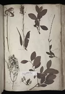
Background imagePlant Anatomy Collection: Lawson plant specimens C016 / 5718

Lawson plant specimens C016 / 5718
Lawson plant specimens. Part of the plant collection attributed to John Lawson, collected in Carolina in 1710. Specimens held in the Sloane Herbarium at the Natural History Museum, London, UK

Background imagePlant Anatomy Collection: Lawson plant specimens C016 / 5717

Lawson plant specimens C016 / 5717
Lawson plant specimens. Part of the plant collection attributed to John Lawson, collected in Carolina in 1710. Specimens held in the Sloane Herbarium at the Natural History Museum, London, UK

Background imagePlant Anatomy Collection: Lawson plant specimens C016 / 5716

Lawson plant specimens C016 / 5716
Lawson plant specimens. Part of the plant collection attributed to John Lawson, collected in Carolina in 1710. Specimens held in the Sloane Herbarium at the Natural History Museum, London, UK

Background imagePlant Anatomy Collection: Himalayan species of Jasmin C016 / 5713

Himalayan species of Jasmin C016 / 5713
Himalayan species of Jasmin. This is the type specimen, the the material used to make the first scientific description of a species

Background imagePlant Anatomy Collection: Amaryllis vittata, artwork C016 / 5710

Amaryllis vittata, artwork C016 / 5710
Amaryllis vittata. Illustration No.3 by Peter Brown, circa 1760s. Held in the Botany Library at the Natural History Museum, London, UK

Background imagePlant Anatomy Collection: Yellow iris (Iris pseudacorus) seeds C016 / 5705

Yellow iris (Iris pseudacorus) seeds C016 / 5705
Yellow iris (Iris pseudacorus) pods and seeds. Many seeds are produced in traingular pods and are dispersed by water

Background imagePlant Anatomy Collection: Prickly pear cactus (Opuntia camachoi) C016 / 5686

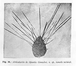
Prickly pear cactus (Opuntia camachoi) C016 / 5686
Prickly pear cactus (Opuntia camachoi). Figure 30 from Revista Chilena de Historia Natural (1932-1933), by Professor Carlos Porter

Background imagePlant Anatomy Collection: Solitary clematis (Clematis integrifolia) C016 / 5688

Solitary clematis (Clematis integrifolia) C016 / 5688
Solitary clematis (Clematis integrifolia). Watercolour by Arthur Harry Church, 10 July 1909

Background imagePlant Anatomy Collection: Barrenwort (Epimedium roseum) C016 / 5689

Barrenwort (Epimedium roseum) C016 / 5689
Barrenwort (Epimedium roseum). Watercolour (Plate 101) by Arthur Harry Church, 9 May 1909

Background imagePlant Anatomy Collection: Monkshood (Aconitum variegatum) C016 / 5690

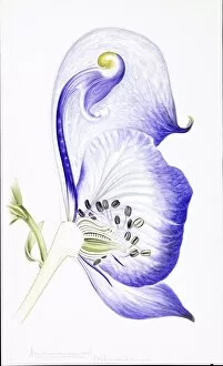
Monkshood (Aconitum variegatum) C016 / 5690
Monkshood (Aconitum variegatum). Watercolour by Arthur Harry Church, 30 July 1906

Background imagePlant Anatomy Collection: Prick nose (Lychnis coronaria) C016 / 5687

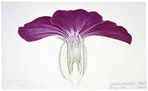
Prick nose (Lychnis coronaria) C016 / 5687
Prick nose (Lychnis coronaria). Watercolour (Plate 171) by Arthur Harry Church, 12 July 1905

Background imagePlant Anatomy Collection: Golden columbine (Aquilegia chrysantha) C016 / 5685

Golden columbine (Aquilegia chrysantha) C016 / 5685
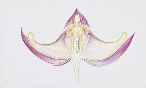
Golden columbine (Aquilegia chrysantha). Ink and wash drawing by Arthur Harry Church, 1906. Figure for Types of floral mechanism. Natural History Museum, London, UK

Background imagePlant Anatomy Collection: Honesty (Lunaria biennis), artwork C016 / 5676

Honesty (Lunaria biennis), artwork C016 / 5676
Hinesty (Lunaria biennis). Watercolour (Plate 146) by Arthur Harry Church. Held in the Botany Library at the Natural History Museum, London, UK

Background imagePlant Anatomy Collection: Franklandia fucifolia, artwork C016 / 5645

Franklandia fucifolia, artwork C016 / 5645
Franklandia fucifolia. Proof etching by I. Pye (based on Ferdinand Bauers drawing) prepared for A voyage to Terra Australis (1814) by Matthew Flinders

Background imagePlant Anatomy Collection: Passion flower (Passiflora aurantia) C016 / 5644

Passion flower (Passiflora aurantia) C016 / 5644
Passion flower (Passiflora aurantia). Engraving by Weddell (based on Ferdinand Bauers unpublished drawing) published in Collectanea botanica (1821) by John Lindley


All Professionally Made to Order for Quick Shipping

"Exploring the Intricate World of Plant Anatomy: A Fascinating Journey through Micrographs and Artwork" Delve into the hidden wonders with these captivating images. From dicotyledon plant stems to castor oil stems, each light micrograph unveils intricate structures that support life. Witness the mesmerizing beauty of Rhynchoglossum obliquum in artwork C016 / 5646, as its delicate details come to life. Dive beneath the surface with a glimpse at maize roots captured under a light microscope. Marvel at the complexity of English oak leaf pores revealed through scanning electron microscopy (SEM), showcasing nature's precision engineering. Take a closer look at the enchanting buttercup flower and tomato leaf, both magnified by light micrography. Explore pollination like never before, as SEM captures this pivotal moment in stunning detail. Uncover secrets hidden within French lavender leaf pores using SEM, revealing their unique structure and function. Witness gutation water droplets on dicot plants' leaf hairs through an unknown magnification SEM image - a phenomenon that showcases nature's ability to adapt and thrive. Embark on an exploration of Ladys mantle reproductive parts with SEM imagery, offering insights into reproduction strategies employed by these remarkable plants. Finally, get up close and personal with primrose pollen under SEM - tiny particles that play a crucial role in plant reproduction. Join us on this visual journey where science meets artistry; discover how every nook and cranny holds mysteries waiting to be unraveled in the fascinating world of plant anatomy.