Greetings Card > Animals > Mammals > Cricetidae > Large Deer Mouse
Greetings Card : Deer antler, SEM


![]()
Cards from Science Photo Library
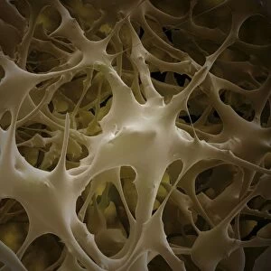
Deer antler, SEM
Deer antler. Coloured scanning electron micrograph (SEM) of a transverse section through cortical (compact) bone from the antler of a deer. The larger spaces seen here are Haversian canals, which contain blood and lymph vessels and nerves. The smaller spaces, known as lacunae, house the osteocytes, the bone-forming cells. Antlers consist entirely of bone and are shed every year. Magnification: x200 when printed at 10 centimetres across
Science Photo Library features Science and Medical images including photos and illustrations
Media ID 9195159
© POWER AND SYRED/SCIENCE PHOTO LIBRARY
Antler Canals Collagen Compact Bone Cortical Bone Deer Haversian Canal Lacuna Lacunae Lamellae Matrix Osteocyte Transverse Section Sectioned
Greetings Card (7"x5")
Bring a touch of the natural world to your correspondence with our captivating selection from the Media Storehouse range of greeting cards. This design features a stunning Coloured Scanning Electron Micrograph (SEM) image of a transverse section through cortical bone from a deer's antler, captured by the Science Photo Library. A unique and thoughtful choice for nature lovers and science enthusiasts alike, these cards are sure to leave a lasting impression.
Folded Greeting Cards (12.5x17.5 cm) have a laminate finish and are supplied with an envelope. The front and inside can be personalised with text in a selection of fonts, layouts and colours.
Greetings Cards suitable for Birthdays, Weddings, Anniversaries, Graduations, Thank You and much more
Estimated Product Size is 17.5cm x 12.5cm (6.9" x 4.9")
These are individually made so all sizes are approximate
Artwork printed orientated as per the preview above, with landscape (horizontal) or portrait (vertical) orientation to match the source image.
EDITORS COMMENTS
This print showcases the intricate beauty of a deer antler, captured through a scanning electron microscope (SEM). The image reveals a transverse section of the antler's cortical bone, providing an up-close look at its remarkable structure. The larger spaces visible in this vibrant SEM are known as Haversian canals. These canals serve as vital passageways for blood vessels, lymph vessels, and nerves within the antler. Meanwhile, the smaller spaces called lacunae house osteocytes - specialized cells responsible for forming new bone tissue. Antlers hold great significance in nature as they are entirely composed of bone and undergo an annual shedding process. This photograph offers us a glimpse into their extraordinary composition and highlights the marvels of biological adaptation. Printed at 10 centimeters across with a magnification of x200, this image from Science Photo Library immerses viewers in the fascinating world of zoology and biology. It serves as a reminder that even seemingly ordinary elements like deer antlers possess incredible complexity when examined closely. With its rich colors and detailed textures, this print is perfect for those who appreciate both artistry and scientific exploration. Whether displayed in homes or educational settings, it invites contemplation on the wonders found within nature's creations.
MADE IN AUSTRALIA
Safe Shipping with 30 Day Money Back Guarantee
FREE PERSONALISATION*
We are proud to offer a range of customisation features including Personalised Captions, Color Filters and Picture Zoom Tools
SECURE PAYMENTS
We happily accept a wide range of payment options so you can pay for the things you need in the way that is most convenient for you
* Options may vary by product and licensing agreement. Zoomed Pictures can be adjusted in the Cart.