Greetings Card : Iris pigment epithelium, SEM


![]()
Cards from Science Photo Library
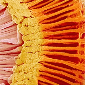
Iris pigment epithelium, SEM
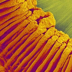
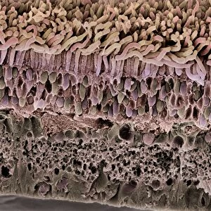
Iris pigment epithelium. Coloured scanning electron micrograph (SEM) of a section through the iris of an eye, showing the iris pigment epithelium (IPE). The IPE is a layer of cuboidal cells (pink) that lies behind the iris. Each cell contains numerous large melanosomes (blue), which contain the pigment melanin. The concentration of this melanin is one of the factors that determine the colour of a persons eye. Magnification: x3, 300 when printed 10 centimetres wide
Science Photo Library features Science and Medical images including photos and illustrations
Media ID 6350495
© STEVE GSCHMEISSNER/SCIENCE PHOTO LIBRARY
Colored Colour Epithelial False Colored Inside Internal Melanin Ocular Ophtalmological Ophthalmology Physiological Physiology Sight Tissue Vision Cells False Coloured Section Sectioned
Greetings Card (7"x5")
Explore the wonders of the human body with our unique range of Science Greetings Cards from Media Storehouse. This eye-catching design features a captivating coloured Scanning Electron Micrograph (SEM) image of the Iris Pigment Epithelium by Steve Gschmeissner from Science Photo Library. Delve into the intricacies of the iris and add a touch of scientific curiosity to your correspondence. Perfect for birthdays, thank yous or just to brighten someone's day, these cards are sure to impress the science enthusiasts in your life. Immerse yourself in the beauty of science and express your thoughts in a thoughtful and visually stunning way. Order yours today and discover the world in a whole new light!
Folded Greeting Cards (12.5x17.5 cm) have a laminate finish and are supplied with an envelope. The front and inside can be personalised with text in a selection of fonts, layouts and colours.
Greetings Cards suitable for Birthdays, Weddings, Anniversaries, Graduations, Thank You and much more
Estimated Product Size is 12.5cm x 17.5cm (4.9" x 6.9")
These are individually made so all sizes are approximate
Artwork printed orientated as per the preview above, with landscape (horizontal) or portrait (vertical) orientation to match the source image.
EDITORS COMMENTS
This print showcases the intricate beauty of the iris pigment epithelium (IPE) found in our eyes. In this coloured scanning electron micrograph, we are granted a glimpse into the hidden world of biology and tissue that contributes to our vision. The IPE, depicted here as a layer of delicate pink cuboidal cells, resides behind the iris and plays a crucial role in determining eye color. Each cell within this layer contains numerous large melanosomes, represented by their striking blue hue. These melanosomes house melanin pigment, which is responsible for giving color to our eyes. With a magnification of x3,300 when printed at 10 centimeters wide, this image allows us to appreciate the astonishing level of detail present in even the smallest components of our bodies. It serves as a reminder that there is so much more than meets the naked eye when it comes to understanding human anatomy and physiology. Photographer Steve Gschmeissner skillfully captures both artistry and scientific precision with his false-colored scanning electron micrograph. This mesmerizing composition not only appeals to those interested in ophthalmology or ocular health but also provides an opportunity for all viewers to marvel at nature's remarkable design within ourselves.
MADE IN AUSTRALIA
Safe Shipping with 30 Day Money Back Guarantee
FREE PERSONALISATION*
We are proud to offer a range of customisation features including Personalised Captions, Color Filters and Picture Zoom Tools
SECURE PAYMENTS
We happily accept a wide range of payment options so you can pay for the things you need in the way that is most convenient for you
* Options may vary by product and licensing agreement. Zoomed Pictures can be adjusted in the Cart.