Jigsaw Puzzle : SEM of Paramecium


![]()
Jigsaw Puzzles from Science Photo Library
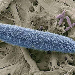
SEM of Paramecium
Paramecium. Coloured scanning electron micrograph (SEM) of the single-celled protozoan, Paramecium caudatum. This ciliate animal has cilia on its cell membrane surface enabling it to swim with an upwards, spiralling motion. It is an elongated oval shape with the groove of its mouth seen at centre. Paramecium is found mainly in stagnant ponds, feeding on bacteria and plant particles. They breed by transverse cell division into two identical cells, while conjugation may occur when two paramecia fuse to facilitate mixing of genetic material. Magnification: x485 at 5x7cm size
Science Photo Library features Science and Medical images including photos and illustrations
Media ID 9307475
© POWER AND SYRED/SCIENCE PHOTO LIBRARY
Ciliate Ellipse Elliptical Micro Organism Mouth Oval Protozoa Protozoan Unicellular
Jigsaw Puzzle (1000 Pieces)
Discover the intricacies of the microscopic world with Media Storehouse's Science-themed Jigsaw Puzzles. Our latest addition, "SEM of Paramecium," showcases this single-celled protozoan in stunning detail. With cilia lining its cell membrane, Paramecium caudatum swims gracefully in water. Assemble this captivating puzzle to bring this coloured scanning electron micrograph (SEM) from Science Photo Library to life and immerse yourself in the fascinating world of science.
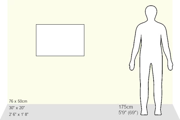
1000 piece puzzles are custom made in Australia and hand-finished on 100% recycled 1.6mm thick laminated puzzle boards. There is a level of repetition in jigsaw shapes with each matching piece away from its pair. The completed puzzle measures 76x50cm and is delivered packaged in an attractive presentation box specially designed to fit most mail slots with a unique magnetic lid
Jigsaw Puzzles are an ideal gift for any occasion
Estimated Product Size is 76cm x 50.2cm (29.9" x 19.8")
These are individually made so all sizes are approximate
Artwork printed orientated as per the preview above, with landscape (horizontal) or portrait (vertical) orientation to match the source image.
EDITORS COMMENTS
This print showcases the intricate beauty of a Paramecium, a single-celled protozoan. The image, captured using a scanning electron microscope (SEM), reveals the vibrant colors and fascinating details of this ciliate animal. With its elongated oval shape and cilia covering its cell membrane surface, the Paramecium propels itself through water with an elegant upward spiraling motion. At the center of this microscopic marvel, we can observe the groove that serves as its mouth. This feature allows the Paramecium to feed on bacteria and plant particles found in stagnant ponds where it predominantly resides. Reproduction for these unicellular organisms occurs through transverse cell division, resulting in two identical cells. However, conjugation is also possible when two paramecia fuse together to facilitate genetic material mixing. Magnified at 485 times its actual size and printed on a 5x7cm medium, this SEM photograph provides us with an extraordinary glimpse into the hidden world of microorganisms. It reminds us of nature's remarkable diversity and complexity even at scales invisible to our naked eyes. This mesmerizing image from Science Photo Library captures not only scientific interest but also sparks curiosity about our natural surroundings. Its vivid portrayal invites contemplation about life's intricacies while highlighting the wonders that exist within every corner of our planet's ecosystems.
MADE IN AUSTRALIA
Safe Shipping with 30 Day Money Back Guarantee
FREE PERSONALISATION*
We are proud to offer a range of customisation features including Personalised Captions, Color Filters and Picture Zoom Tools
SECURE PAYMENTS
We happily accept a wide range of payment options so you can pay for the things you need in the way that is most convenient for you
* Options may vary by product and licensing agreement. Zoomed Pictures can be adjusted in the Cart.