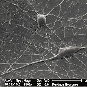
Metal Print : Purkinje nerve cell, SEM


![]()
Metal Prints from Science Photo Library
Purkinje nerve cell, SEM
Science Photo Library features Science and Medical images including photos and illustrations
Media ID 6421882
© DAVID MCCARTHY/SCIENCE PHOTO LIBRARY
Brown Cerebellum Connection Connections Dendrite Dendrites Granular Grey Matter Histology Junction Molecular Layer Nerve Cell Nervous Neuron Neurone Process Processes Purkinje System Tissue Brain Cells Neurology
14"x11" (28x35cm) Metal Print
Discover the intricacy of nature with our Media Storehouse Metal Prints featuring the Purkinje Nerve Cell, captured in stunning detail through Science Photo Library's SEM image. Each print is meticulously crafted using high-quality metal materials, ensuring vibrant colors and exceptional clarity. Bring the beauty of science into your home or office space and ignite curiosity with this captivating representation of the complex Purkinje Nerve Cell.
Our Metal Prints feature rounded corners and rear fixings for easy wall mounting. Images are directly printed onto a lightweight, high quality 5mm thick, durable metallic surface for a vivid and vibrant finish. Available in 2 sizes, 28x35cm (14x11) and 50x40cm (20x16). The unique material is fade, moisture, chemical and scratch resistant to help ensure this art lasts a lifetime.
Made with durable metal and luxurious printing techniques, metal prints bring images to life and add a modern touch to any space
Estimated Product Size is 35.5cm x 27.9cm (14" x 11")
These are individually made so all sizes are approximate
Artwork printed orientated as per the preview above, with landscape (horizontal) or portrait (vertical) orientation to match the source image.
EDITORS COMMENTS
This print showcases the intricate beauty of a Purkinje nerve cell, captured using scanning electron microscopy (SEM). The image reveals the complex network of connections and processes that make up this vital component of our nervous system. In vibrant colors, we see the brown and white branches representing dendrites, which extend from the main body of the neuron known as the soma. These dendrites play a crucial role in receiving signals from other neurons and transmitting them to the soma. The molecular layer surrounding this Purkinje cell is depicted in shades of grey, highlighting its position within the cerebellum - an area responsible for motor control and coordination. This region is rich in granular cells, which can be seen forming connections with multiple dendrites. The detailed structure visible in this SEM image emphasizes both the complexity and elegance inherent in our neural architecture. It serves as a reminder of how essential these microscopic components are for maintaining a healthy functioning brain. As an invaluable tool for neurology research and histology studies, SEM allows us to explore these fascinating aspects of human anatomy on a cellular level. Science Photo Library has expertly captured this stunning representation that not only educates but also inspires awe at nature's design.
MADE IN AUSTRALIA
Safe Shipping with 30 Day Money Back Guarantee
FREE PERSONALISATION*
We are proud to offer a range of customisation features including Personalised Captions, Color Filters and Picture Zoom Tools
SECURE PAYMENTS
We happily accept a wide range of payment options so you can pay for the things you need in the way that is most convenient for you
* Options may vary by product and licensing agreement. Zoomed Pictures can be adjusted in the Cart.