Mouse Mat > Popular Themes > Human Body
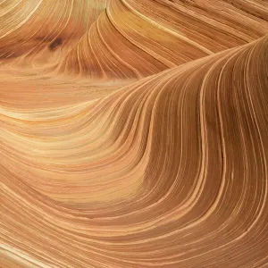
Mouse Mat : Muscle fibre structure, artwork

![]()
Home Decor from Science Photo Library
Muscle fibre structure, artwork
Muscle fibre. Computer artwork showing the structure of a muscle cell, also known as a muscle fibre. The cell is surrounded by a plasma membrane called the sarcolemma (cream). Within the cell are myofibrils, bundles of actin and myosin fibres (pink) surrounded by a sarcoplasmic reticulum (yellow). The sarcoplasmic reticulum holds a reserve of calcium ions, which are needed for contraction. Mitochondria (blue) within the myofibrils provide the energy needed for contraction
Science Photo Library features Science and Medical images including photos and illustrations
Media ID 9242697
© HENNING DALHOFF / SCIENCE PHOTO LIBRARY
Actin Cut Away Fiber Filament Mitochondria Mitochondrion Muscle Fibre Myofibril Myofibrils Myosin Plasma Membrane Sarcolemma Cutouts Muscle Cell Musculature
Mouse Pad
Bring some life into your office, or create a heartfelt gift, with a personalised deluxe Mouse Mat. Made of high-density black foam with a tough, stain-resistant inter-woven cloth cover they will brighten up any home or corporate office.
Archive quality photographic print in a durable wipe clean mouse mat with non slip backing. Works with all computer mice
Estimated Product Size is 24.2cm x 19.7cm (9.5" x 7.8")
These are individually made so all sizes are approximate
Artwork printed orientated as per the preview above, with landscape (horizontal) or portrait (vertical) orientation to match the source image.
EDITORS COMMENTS
This artwork, titled "Muscle Fibre Structure" takes us on a mesmerizing journey into the intricate world of muscle cells. The computer-generated image showcases the remarkable complexity and beauty hidden within our bodies. At first glance, we are drawn to the central muscle cell enveloped by a creamy plasma membrane known as the sarcolemma. Delving deeper, our eyes are captivated by bundles of pink actin and myosin fibers called myofibrils that reside within the cell. These fibrous structures play a crucial role in muscle contraction, allowing us to perform various physical activities. The yellow sarcoplasmic reticulum surrounding the myofibrils catches our attention next. This specialized network acts as a reservoir for calcium ions essential for muscle contractions - an intriguing detail that highlights nature's ingenious design. Within each myofibril lies blue-colored mitochondria, powerhouses responsible for generating energy required during muscular movements. Their presence reminds us of how intricately interconnected every component is within our body. Against a pristine white background, this illustration serves as both an educational tool and an awe-inspiring work of art. It invites viewers to appreciate not only the structural marvels but also gain insight into biology and anatomy. "Muscle Fibre Structure" offers a glimpse into one aspect of human physiology while reminding us of the wonders concealed beneath our skin's surface – an exquisite testament to life's complexity captured through scientific artistry.
MADE IN AUSTRALIA
Safe Shipping with 30 Day Money Back Guarantee
FREE PERSONALISATION*
We are proud to offer a range of customisation features including Personalised Captions, Color Filters and Picture Zoom Tools
SECURE PAYMENTS
We happily accept a wide range of payment options so you can pay for the things you need in the way that is most convenient for you
* Options may vary by product and licensing agreement. Zoomed Pictures can be adjusted in the Cart.