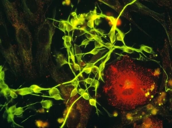
Immunofluorescent LM of macrophage in brain tissue
![]()

Wall Art and Photo Gifts from Science Photo Library
Immunofluorescent LM of macrophage in brain tissue
Immunofluorescent Light Micrograph of a macrophage within brain tissue. Macrophages found in nervous tissue are termed microglia. At lower right, this large scavenger cell (stained red) moves in search of bacteria and other foreign invaders to ingest, playing an important role in the bodys immune response. At centre, a tangle of neuron cells with interconnected nerve fibres stains green. Impulses travel through this tissue. Other non-conducting cells (glia) which form a supportive network, are also seen. Immunofluorescence is a staining technique which uses antibodies to attach fluores- cent dyes to specific tissues and molecules in the cell. Magnification x400 at 35mm, x750 at 6x4.5cm
Science Photo Library features Science and Medical images including photos and illustrations
Media ID 6421224
© NANCY KEDERSHA/UCLA/SCIENCE PHOTO LIBRARY
Active Defence Immune Immunofluores Immunology Macrophage Microglia Phagocyte System Brain Cells Light Micrograph
EDITORS COMMENTS
This print from Science Photo Library showcases the intricate world of the human immune system within brain tissue. The image, captured using immunofluorescent light microscopy, reveals a macrophage - a type of phagocyte - in vibrant red as it diligently searches for bacteria and other foreign invaders to engulf and eliminate. This large scavenger cell plays a crucial role in our body's defense mechanism. At the center of the composition, we witness a mesmerizing tangle of neuron cells with their interconnected nerve fibers glowing in vivid green. These cells are responsible for transmitting electrical impulses throughout this vital tissue network. Surrounding them are non-conducting glial cells that form an essential supportive network. Immunofluorescence, an advanced staining technique employing fluorescent dyes attached to specific tissues and molecules within cells through antibodies, allows us to visualize these microscopic wonders with astonishing clarity. With magnification at x400 on 35mm film or x750 on 6x4.5cm format, this image offers us a glimpse into the complex anatomy of our immune system within the brain. Through this remarkable visual representation, we gain insight into how our bodies actively defend against harmful pathogens while maintaining delicate neural connections critical for proper functioning. Science Photo Library continues to provide awe-inspiring scientific images that bridge artistry and knowledge acquisition without compromising commercial use restrictions
MADE IN AUSTRALIA
Safe Shipping with 30 Day Money Back Guarantee
FREE PERSONALISATION*
We are proud to offer a range of customisation features including Personalised Captions, Color Filters and Picture Zoom Tools
SECURE PAYMENTS
We happily accept a wide range of payment options so you can pay for the things you need in the way that is most convenient for you
* Options may vary by product and licensing agreement. Zoomed Pictures can be adjusted in the Cart.

