Home > Arts > Street art graffiti > Digital art > Digital paintings
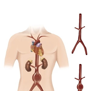
Ureterovesical junction (UVJ) in the kidney
 in the kidney")
![]()
Wall Art and Photo Gifts from Stocktrek
Ureterovesical junction (UVJ) in the kidney
Ureterovesical junction (UVJ) in the kidney. This is the point where the ureters from the kidney connect to the bladder
Stocktrek Images specializes in Astronomy, Dinosaurs, Medical, Military Forces, Ocean Life, & Sci-Fi
Media ID 13008003
© Stocktrek Images
Abnormal Anatomy Artery Biology Biomedical Illustrations Blood Flow Colored Background Cross Section Cutaway View Digestion Digestive System Filter Gland Healthcare Human Anatomy Human Body Parts Human Glands Human Kidneys Human Organs Interlobular Artery Internal Organs Kidney Major Calyx Medical Medicine Medulla Minor Calyx Nephrology Nephron Organ Physiology Renal Artery Renal Capsule Renal Circulation Renal Column Renal Hilum Renal Papilla Renal Pelvis Renal Pyramids Swollen Ureter Urinary System Kidney Stones Urolithiasis
FEATURES IN THESE COLLECTIONS
> Arts
> Street art graffiti
> Digital art
> Digital paintings
> Posters
> Aircraft Posters
> Cutaway Posters
EDITORS COMMENTS
This print from Stocktrek Images showcases the intricate Ureterovesical junction (UVJ) in the kidney. The UVJ serves as a vital connection point where the ureters, responsible for carrying urine from the kidneys, merge with the bladder. The image features a cross-section of this remarkable anatomical structure against a colored background, providing an excellent visual representation of its complexity. It highlights various components such as arteries, glands, and renal pyramids that contribute to its functionality within our bodies. With its detailed depiction of internal organs and biomedical illustrations, this photograph is not only visually appealing but also holds significant medical value. It offers insights into nephrology and renal circulation while shedding light on conditions like hydronephrosis and urolithiasis (kidney stones). The cutaway view allows us to observe blood flow through interlobular arteries and witness how urine is filtered through nephrons before being transported via ureters to the bladder. This comprehensive understanding of human anatomy aids healthcare professionals in diagnosing abnormalities or complications related to urinary system functioning. Whether you are a biology enthusiast or a medical practitioner seeking educational resources, this single object image provides an invaluable tool for studying human glands and gaining insight into one's overall health.
MADE IN AUSTRALIA
Safe Shipping with 30 Day Money Back Guarantee
FREE PERSONALISATION*
We are proud to offer a range of customisation features including Personalised Captions, Color Filters and Picture Zoom Tools
SECURE PAYMENTS
We happily accept a wide range of payment options so you can pay for the things you need in the way that is most convenient for you
* Options may vary by product and licensing agreement. Zoomed Pictures can be adjusted in the Cart.