Framed Print : Diagram of cell wall & flagellum of Gram- bacteria


![]()
Framed Photos from Science Photo Library
Diagram of cell wall & flagellum of Gram- bacteria
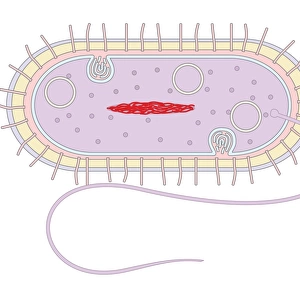
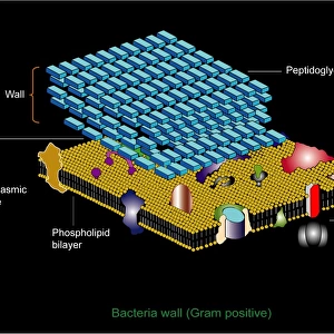
Diagrammatic representation of the cell wall, cell membrane, and a flagellum of a Gram-negative bacterium. Gram-negative bacteria have an outer lipopolysaccharide layer (above, yellow), as an outer membrane to the cell wall. Beneath it is the peptidoglycan cell wall, rigid & protective, made of sheets of glycan (sugar, white bricks) with peptide cross-links. The true cell membrane is at bottom (yellow, phospholipid). Pathogens attach to both membranes, molecules diffuse through, & anti- gens are found on them. The rotor mechanism of a flagellum is portrayed (red and blue); it rotates the flagellum like a propeller for movement
Science Photo Library features Science and Medical images including photos and illustrations
Media ID 6400531
© FRANCIS LEROY, BIOCOSMOS/SCIENCE PHOTO LIBRARY
Bacteria Cell Membrane Cell Structure Cell Wall Cytology Flagellum Gram Negative Micro Biology
13.5"x11.5" (34x29cm) Premium Frame
Bring the fascinating world of microbiology into your home or office with our Framed Prints from Media Storehouse. This captivating diagram, sourced from Science Photo Library, illustrates the complex structure of a Gram-negative bacterium, featuring a clear depiction of its cell wall, cell membrane, and a flagellum. The intricate details of this diagram, including the outer lipopolysaccharide layer, are sure to impress and inspire. Each print is carefully framed to preserve and enhance the image, making it a beautiful addition to any space.
Framed and mounted 9x7 print. Professionally handmade full timber moulded frames are finished off with framers tape and come with a hanging solution on the back. Outer dimensions are 13.5x11.5 inches (34x29cm). Quality timber frame frame moulding (20mm wide and 30mm deep) with frame colours in your choice of black, white, or raw oak and a choice of black or white card mounts. Frames have a perspex front providing a virtually unbreakable glass-like finish which is easily cleaned with a damp cloth.
Contemporary Framed and Mounted Prints - Professionally Made and Ready to Hang
Estimated Image Size (if not cropped) is 21.4cm x 21.4cm (8.4" x 8.4")
Estimated Product Size is 34cm x 29.2cm (13.4" x 11.5")
These are individually made so all sizes are approximate
Artwork printed orientated as per the preview above, with landscape (horizontal) or portrait (vertical) orientation to match the source image.
EDITORS COMMENTS
This print showcases a detailed diagrammatic representation of the cell wall, cell membrane, and flagellum of a Gram-negative bacterium. The complexity and intricacy of these microscopic structures are beautifully captured in this artwork. The outer lipopolysaccharide layer, depicted in vibrant yellow above the diagram, forms an outer membrane to the cell wall. This layer acts as a protective shield for the bacterium. Beneath it lies the peptidoglycan cell wall, portrayed as rigid white bricks with peptide cross-links. This structure provides further protection and support to the bacterium. At the bottom of the diagram is the true cell membrane, represented by a striking yellow coloration due to its phospholipid composition. Both membranes serve as attachment points for pathogens while allowing molecules to diffuse through them. Additionally, antigens can be found on these membranes. Highlighted in red and blue is the rotor mechanism of a flagellum – an appendage that enables movement like a propeller. Its presence emphasizes how bacteria utilize this intricate structure for mobility. This artful depiction not only captures essential elements of cytology but also serves as an educational tool for understanding bacterial biology at its core. It reminds us of both their remarkable structural complexity and their role in various biological processes.
MADE IN AUSTRALIA
Safe Shipping with 30 Day Money Back Guarantee
FREE PERSONALISATION*
We are proud to offer a range of customisation features including Personalised Captions, Color Filters and Picture Zoom Tools
SECURE PAYMENTS
We happily accept a wide range of payment options so you can pay for the things you need in the way that is most convenient for you
* Options may vary by product and licensing agreement. Zoomed Pictures can be adjusted in the Cart.