Fraud Blocker Skip to main content

Biochemistry Collection (page 7)

Biochemistry is the fascinating study of the chemical processes and substances that occur within living organisms

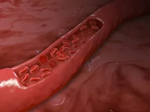
Background imageBiochemistry Collection: Artery cross section with red blood cell flow

Artery cross section with red blood cell flow

Background imageBiochemistry Collection: Conceptual image of lysosome

Conceptual image of lysosome. Lysosomes are cellular organelles that contain acid hydrolase enzymes that break down waste materials and cellular debris

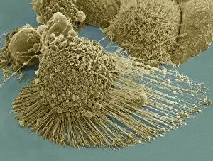
Background imageBiochemistry Collection: Scanning electron micrograph of an apoptotic HeLa cell

Scanning electron micrograph of an apoptotic HeLa cell. Zeiss Merlin HR-SEM

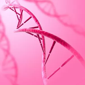
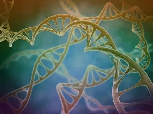
Background imageBiochemistry Collection: Conceptual image of DNA

Conceptual image of DNA

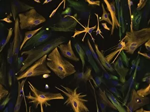
Background imageBiochemistry Collection: Directed differentiation of multipotential human neural progenitor cells

Directed differentiation of multipotential human neural progenitor cells
Human neural progenitor cells were isolated under selective culture conditions from the developing human brain and directed through lineage differentiation to GFAP + (glial fibrillary acid protein)

Background imageBiochemistry Collection: Microscopic view of HIV virus

Microscopic view of HIV virus

Background imageBiochemistry Collection: Cluster of DNA strands

Cluster of DNA strands of human DNA or deoxyribonucleic acid

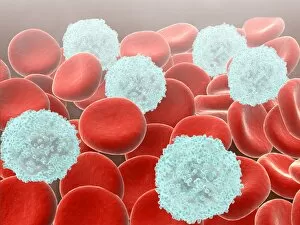
Background imageBiochemistry Collection: Red blood cells with white blood cells

Red blood cells with white blood cells

Background imageBiochemistry Collection: Conceptual image of common bacteria

Conceptual image of common bacteria

Background imageBiochemistry Collection: Illustration of a Brontosaurus chemist

Illustration of a Brontosaurus chemist

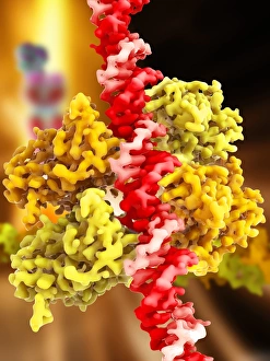
Background imageBiochemistry Collection: DNA transcription, illustration C018 / 0900

DNA transcription, illustration C018 / 0900
DNA (deoxyribonucleic acid) transcription. Illustration of an RNA (ribonucelic acid) polymerase molecule (centre) synthesising an mRNA (messenger RNA) strand (bottom)

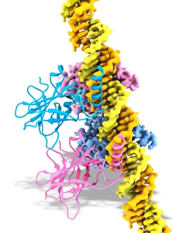
Background imageBiochemistry Collection: Tumour suppressor protein and DNA C017 / 3647

Tumour suppressor protein and DNA C017 / 3647
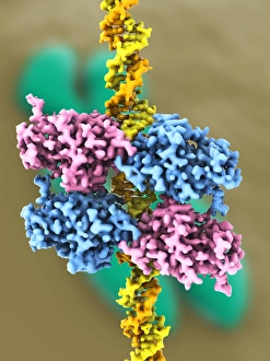
Tumour suppressor protein and DNA. Computer artwork showing a molecule of the tumour suppressor protein p53 (blue and pink) bound to a molecule of DNA (deoxyribonucleic acid, yellow and orange)

Background imageBiochemistry Collection: Ricin A-chain, artwork C017 / 3653

Ricin A-chain, artwork C017 / 3653
Ricin A-chain. Computer artwork showing the enzymatically active A-chain from a molecule of the toxic protein ricin. Ricin comprises two entwined amino acid chains; A (seen here) and B (not shown)

Background imageBiochemistry Collection: TATA box-binding protein complex C017 / 7082

TATA box-binding protein complex C017 / 7082
TATA box-binding protein complex. Molecular model showing a TATA box-binding protein (TBP, green) complexed with a strand of DNA (deoxyribonucleic acid, yellow) and transcription factor IIB

Background imageBiochemistry Collection: Antibiotic resistance enzyme molecule C017 / 2272

Antibiotic resistance enzyme molecule C017 / 2272
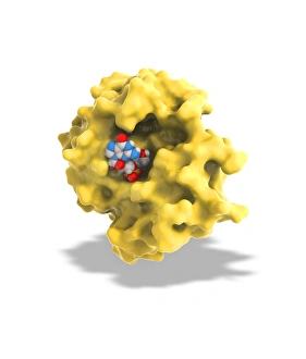
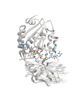
Antibiotic resistance enzyme. Molecular model of the New Delhi metallo-beta-lactamase 1 enzyme. This bacterial enzyme confers antibiotic resistance on cells that carry it

Background imageBiochemistry Collection: DNA molecule, artwork C017 / 7217

DNA molecule, artwork C017 / 7217
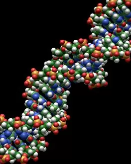
DNA molecule. Computer artwork showing a double stranded DNA (deoxyribonucleic acid) molecule. DNA is composed of two strands twisted into a double helix

Background imageBiochemistry Collection: DNA molecule, artwork C017 / 0616

DNA molecule, artwork C017 / 0616
DNA molecule. Computer artwork looking along the interior of a double stranded DNA (deoxyribonucleic acid) molecule. DNA is composed of two strands twisted into a double helix

Background imageBiochemistry Collection: TATA box-binding protein complex C017 / 7088

TATA box-binding protein complex C017 / 7088
TATA box-binding protein complex. Molecular model showing a TATA box-binding protein (TBP, green) complexed with a strand of DNA (deoxyribonucleic acid, yellow) and transcription factor IIB

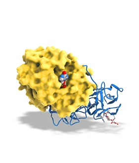
Background imageBiochemistry Collection: Ricin molecule, artwork C017 / 3652

Ricin molecule, artwork C017 / 3652
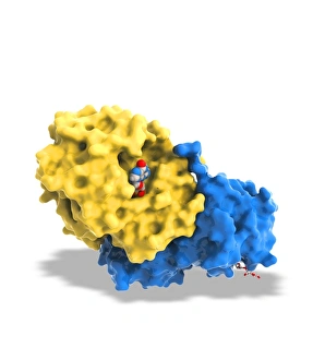
Ricin molecule. Computer artwork showing the structure of a molecule of the toxic protein ricin. Ricin comprises two entwined amino acid chains; A (yellow) and B (blue)

Background imageBiochemistry Collection: DNA molecule, artwork C017 / 0615

DNA molecule, artwork C017 / 0615
DNA molecule. Computer artwork looking along the interior of a double stranded DNA (deoxyribonucleic acid) molecule. DNA is composed of two strands twisted into a double helix

Background imageBiochemistry Collection: DNA molecule, artwork C017 / 0617

DNA molecule, artwork C017 / 0617
DNA molecule. Computer artwork looking along the interior of a double stranded DNA (deoxyribonucleic acid) molecule. DNA is composed of two strands twisted into a double helix

Background imageBiochemistry Collection: Antibiotic resistance enzyme molecule C017 / 2271

Antibiotic resistance enzyme molecule C017 / 2271
Antibiotic resistance enzyme. Molecular model of the New Delhi metallo-beta-lactamase 1 enzyme. This bacterial enzyme confers antibiotic resistance on cells that carry it

Background imageBiochemistry Collection: TATA box-binding protein complex C017 / 7084

TATA box-binding protein complex C017 / 7084
TATA box-binding protein complex. Molecular model showing a TATA box-binding protein (TBP, green) complexed with a strand of DNA (deoxyribonucleic acid, yellow) and transcription factor IIB

Background imageBiochemistry Collection: Sirtuin enzyme and p53, artwork C017 / 3659

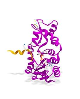
Sirtuin enzyme and p53, artwork C017 / 3659
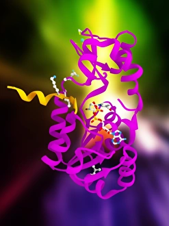
Sirtuin enzyme and p53. Computer artwork of a sirtuin (Sir2) enzyme (pink) bound to a p53 peptide (orange). Sir2 enzymes form a unique class of NAD(+)

Background imageBiochemistry Collection: Adenine molecule, artwork C017 / 7200

Adenine molecule, artwork C017 / 7200
Adenine molecule. Computer artwork showing the structure of a molecule of the nucleobase adenine. Atoms are colour-coded spheres: carbon (green), nitrogen (blue), and oxygen (white)
Background imageBiochemistry Collection: Tumour suppressor protein and DNA C017 / 3644

Tumour suppressor protein and DNA C017 / 3644
Tumour suppressor protein and DNA. Computer artwork showing a molecule of the tumour suppressor protein p53 (blue and pink) bound to a molecule of DNA (deoxyribonucleic acid, yellow and orange)

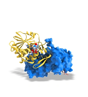
Background imageBiochemistry Collection: Ricin molecule, artwork C017 / 3651

Ricin molecule, artwork C017 / 3651
Ricin molecule. Computer artwork showing the structure of a molecule of the toxic protein ricin. Ricin comprises two entwined amino acid chains; A (yellow) and B (blue)

Background imageBiochemistry Collection: Ricin molecule, artwork C017 / 3650

Ricin molecule, artwork C017 / 3650
Ricin molecule. Computer artwork showing the structure of a molecule of the toxic protein ricin. Ricin comprises two entwined amino acid chains; A (yellow) and B (blue)

Background imageBiochemistry Collection: Sirtuin enzyme and p53, artwork C017 / 3658

Sirtuin enzyme and p53, artwork C017 / 3658
Sirtuin enzyme and p53. Computer artwork of a sirtuin (Sir2) enzyme (pink) bound to a p53 peptide (orange). Sir2 enzymes form a unique class of NAD(+)

Background imageBiochemistry Collection: Cytosine-guanine interaction, artwork C017 / 7215

Cytosine-guanine interaction, artwork C017 / 7215
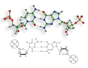
Cytosine-guanine interaction. Computer artwork showing the structure of bound cytosine (left) and guanine molecules (right)

Background imageBiochemistry Collection: SIRT3 molecule, artwork C017 / 3657

SIRT3 molecule, artwork C017 / 3657
SIRT3 molecule. Computer artwork showing the structure of a molecule of NAD-dependent deacetylase sirtuin-3, mitochondrial (SIRT3)

Background imageBiochemistry Collection: Tumour suppressor protein and DNA C017 / 3646

Tumour suppressor protein and DNA C017 / 3646
Tumour suppressor protein and DNA. Computer artwork showing a molecule of the tumour suppressor protein p53 (blue and pink) bound to a molecule of DNA (deoxyribonucleic acid, yellow and orange)

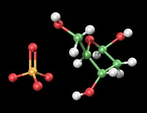
Background imageBiochemistry Collection: DNA components, artwork C017 / 7350

DNA components, artwork C017 / 7350
DNA components. Computer artwork showing the structure of the two molecules that make up the backbone of DNA (deoxyribonucleic acid), phosphate (left) and deoxyribose (right)

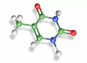
Background imageBiochemistry Collection: Thymine molecule, artwork C017 / 7366

Thymine molecule, artwork C017 / 7366
Thymine molecule. Computer artwork showing the structure of a molecule of the nucleobase thymine. Atoms are colour-coded spheres: carbon (green), nitrogen (blue), oxygen (red), and hydrogen (white)

Background imageBiochemistry Collection: Photosynthesis, artwork C017 / 0788

Photosynthesis, artwork C017 / 0788
Photosynthesis. Conceptual computer artwork of photosynthesis in a plant cell. Photosynthesis is the process by which most plants convert sunlight (coming from top left) into chemical energy

Background imageBiochemistry Collection: Thymine molecule, artwork C017 / 7365

Thymine molecule, artwork C017 / 7365
Thymine molecule. Computer artwork showing the structure of a molecule of the nucleobase thymine. Atoms are colour-coded spheres: carbon (green), nitrogen (blue), oxygen (red), and hydrogen (white)

Background imageBiochemistry Collection: Cytosine-guanine interaction, artwork C017 / 7216

Cytosine-guanine interaction, artwork C017 / 7216
Cytosine-guanine interaction. Computer artwork showing the structure of bound cytosine (left) and guanine molecules (right)

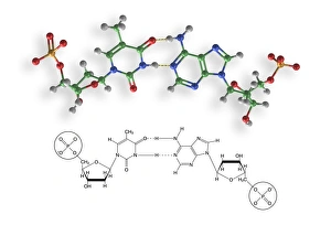
Background imageBiochemistry Collection: Thymine-adenine interaction, artwork C017 / 7367

Thymine-adenine interaction, artwork C017 / 7367
Thymine-adenine interaction. Computer artwork showing the structure of bound thymine and adenine molecules. Atoms are shown as colour-coded spheres: carbon (green), hydrogen (white)

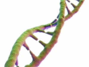
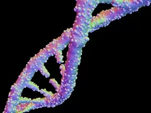
Background imageBiochemistry Collection: DNA molecule, artwork F007 / 4200

DNA molecule, artwork F007 / 4200
DNA molecule, computer artwork

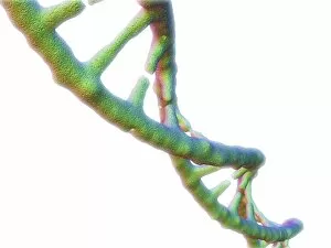
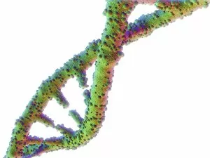
Background imageBiochemistry Collection: DNA molecule, artwork F007 / 4196

DNA molecule, artwork F007 / 4196
DNA molecule, computer artwork

Background imageBiochemistry Collection: DNA molecule, artwork F007 / 4203

DNA molecule, artwork F007 / 4203
DNA molecule, computer artwork

Background imageBiochemistry Collection: DNA molecule, artwork F007 / 4207

DNA molecule, artwork F007 / 4207
DNA molecule, computer artwork

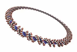
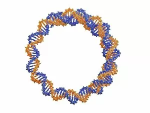
Background imageBiochemistry Collection: Circular DNA molecule, artwork F006 / 7088

Circular DNA molecule, artwork F006 / 7088
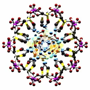
Circular DNA (deoxyribonucleic acid) molecule, computer artwork. Circular DNA has no ends, but consists of a ring structure

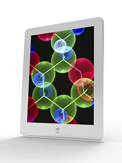
Background imageBiochemistry Collection: Tablet computer showing a DNA molecule F006 / 6310

Tablet computer showing a DNA molecule F006 / 6310
Tablet computer showing artwork of a DNA molecule

Background imageBiochemistry Collection: Circular DNA molecule, space artwork F006 / 7089

Circular DNA molecule, space artwork F006 / 7089
Circular DNA (deoxyribonucleic acid) molecule, computer artwork and space nebula artwork, depicting origin of life

Background imageBiochemistry Collection: Tablet computer, insulin molecule F006 / 6311

Tablet computer, insulin molecule F006 / 6311
Tablet computer showing a part of the molecule of human insulin. A single insulin molecule is made up of two chains of amino acids, the A and B chains, which are held together by di-sulphide bridges

Background imageBiochemistry Collection: Circular DNA molecule, artwork F006 / 7072

Circular DNA molecule, artwork F006 / 7072
Circular DNA (deoxyribonucleic acid) molecule, computer artwork. Circular DNA has no ends, but consists of a ring structure

Background imageBiochemistry Collection: DNA molecule, artwork F006 / 3715

DNA molecule, artwork F006 / 3715
DNA molecule, computer artwork


All Professionally Made to Order for Quick Shipping

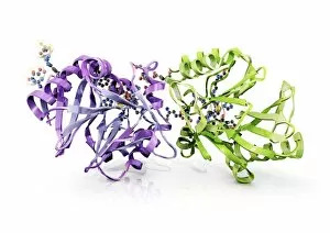
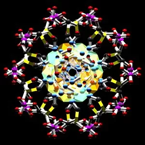
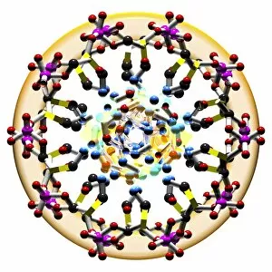
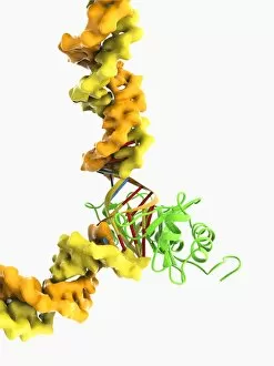
Biochemistry is the fascinating study of the chemical processes and substances that occur within living organisms. In this captivating field, scientists delve into the intricate mechanisms that govern life itself. One intriguing aspect involves anaesthetics inhibiting an ion channel (C015 / 6718), revealing how these compounds can block nerve signals and induce temporary loss of sensation. Another captivating image showcases the structure of an Immunoglobulin G antibody molecule, highlighting its crucial role in our immune system's defense against pathogens. In a mesmerizing light micrograph, EDTA crystals are captured in all their splendor, showcasing their unique geometric patterns. Similarly, another image displays Immunoglobulin G antibody molecules (F007 / 9894) with astonishing detail, emphasizing their ability to recognize and neutralize foreign invaders. The beauty extends beyond static images; it also encompasses dynamic processes. A stunning artwork depicts the cell membrane (C013 / 7467), illustrating its vital function as a barrier and gatekeeper for essential molecules entering or leaving cells. Exploring further into our body's inner workings, we encounter a captivating artwork depicting the blood coagulation cascade (C016 / 9873). This complex series of reactions ensures proper clotting when injuries occur to prevent excessive bleeding. DNA takes center stage in another illustration, showcasing its double helix structure that carries genetic information responsible for traits passed down from generation to generation. Additionally, a microscopic view reveals human respiratory syncytial virus—a reminder of both the beauty and danger found at microscopic levels. Delving deeper into molecular interactions, we witness zinc fingers bound to a DNA strand—an elegant representation of how proteins regulate gene expression by binding specific sequences on DNA molecules. It also sheds light on hormones' crystalline forms—oxytocin hormone crystals (PLM C016 / 7196) demonstrate this phenomenon beautifully while testosterone crystals captivate us under polarized light microscopy (PLM).