Fraud Blocker Skip to main content

Cell Collection (page 2)

"Exploring the Intricacies of Life: Unveiling the Wonders of Cells" Delving into the microscopic world

Background imageCell Collection: Stem cells, SEM

Stem cells, SEM
Stem cells, coloured scanning electron micrograph (SEM). Stem cells can differentiate into any other cell type. There are three main types of mammalian stem cell: embryonic stem cells

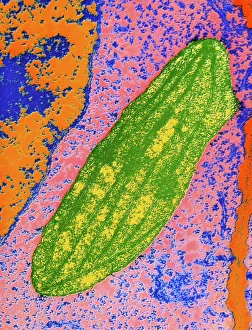
Background imageCell Collection: Chloroplast in cell of pea plant

Chloroplast in cell of pea plant
Coloured transmisson electron micrograph of a chloroplast (green) sitting in the cytoplasm of a pea plant Pisum sativum. The chloroplast is the site of photosynthesis where carbohydrates are obtained

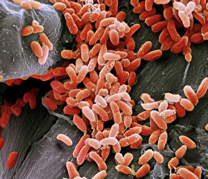
Background imageCell Collection: Pseudomonas aeruginosa bacteria, SEM

Pseudomonas aeruginosa bacteria, SEM
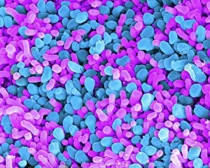
Pseudomonas aeruginosa bacteria, coloured scanning electron micrograph (SEM). These Gram-negative rod-shaped bacteria are found in soil and water, and as normal flora in the human intestine

Background imageCell Collection: Cerebellum tissue, light micrograph

Cerebellum tissue, light micrograph
Cerebellum tissue. Confocal light micrograph of a section through the cerebellum of the brain showing two types of glial cells (support cells); astrocytes (star-shaped)

Background imageCell Collection: Plasmodium sp. malarial parasite

Plasmodium sp. malarial parasite
Scanning electron microscope image of a malarial protozoal parasite. The parasite requires the anopheles mosquito to complete its life cycle

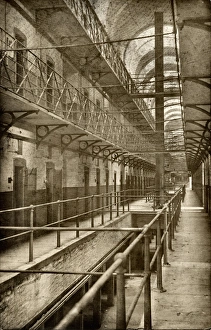
Background imageCell Collection: Cell Block Interior, Wakefield Prison, West Yorkshire

Cell Block Interior, Wakefield Prison, West Yorkshire
The interior of a cell block at Wakefield Prison, West Yorkshire. Cells line the open galleries which have a safety net strung across to prevent inmates falling

Background imageCell Collection: Guillotine

Guillotine
Petri Damstén

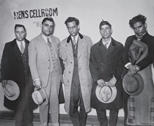
Background imageCell Collection: Joe Aiello (1891-1930) with his henchmen in police custody, 1928 (b / w photo)

Joe Aiello (1891-1930) with his henchmen in police custody, 1928 (b / w photo)
PNP252300 Joe Aiello (1891-1930) with his henchmen in police custody, 1928 (b/w photo) by American Photographer, (20th century); Private Collection; (add.info)

Background imageCell Collection: Solar panel with sun

Solar panel with sun

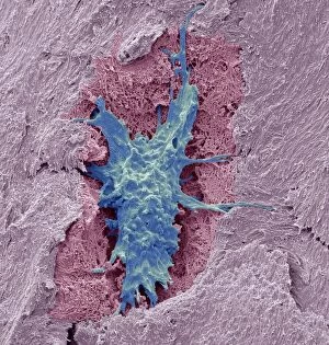
Background imageCell Collection: Osteocyte bone cell, SEM C016 / 9025

Osteocyte bone cell, SEM C016 / 9025
Osteocyte bone cell. Coloured scanning electron micrograph (SEM) of an osteocyte bone cell (blue) surrounded by bone tissue (pink)

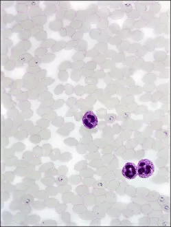
Background imageCell Collection: Dohle bodies in blood cell, micrograph

Dohle bodies in blood cell, micrograph
Dohle bodies in blood cell. Light micrograph of a neutrophil white blood cell (centre) with Dohle bodies. These are small inclusions within the cells cytoplasm
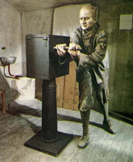
Background imageCell Collection: Convict in prison cell turning hand-crank

Convict in prison cell turning hand-crank

Background imageCell Collection: Top of mans head visible behind prison cell window as his hands grip metal bars, front view

Top of mans head visible behind prison cell window as his hands grip metal bars, front view

Background imageCell Collection: Picture No. 11675528

Picture No. 11675528
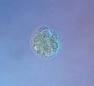
Mouse Embryo at Eight Cell Stage. Date:

Background imageCell Collection: Picture No. 11675527

Picture No. 11675527
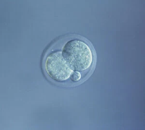
Mouse Embryo at Four Cell Stage & a Polar Body. Date:

Background imageCell Collection: Picture No. 11675526

Picture No. 11675526
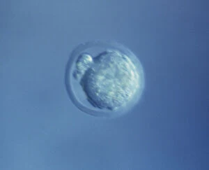
Mouse Embryo at Two Cell Stage & a Polar Body. Date:

Background imageCell Collection: Picture No. 11675525

Picture No. 11675525
Mouse Embryo at Single Cell Stage and Polar Body. Date:

Background imageCell Collection: Picture No. 11675522

Picture No. 11675522
A human Ovum surrounded by Sperm - being fertilized. Date:

Background imageCell Collection: Picture No. 11675612

Picture No. 11675612
Scanning Electron Micrograph (SEM): Human White and Red Blood Cells. Date:

Background imageCell Collection: Picture No. 10876997

Picture No. 10876997
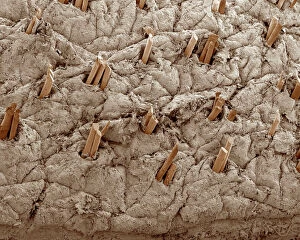
Scanning Electron Micrograph (SEM): Human Skin with Hair Follicles Date:

Background imageCell Collection: Fruit-powered clock

Fruit-powered clock. In this experiment each apple is a voltaic cell. The apples have been connected to make a battery. Copper (orange) and zinc (silver)

Background imageCell Collection: Pancreas anatomy, artwork

Pancreas anatomy, artwork
Pancreas anatomy. Cut-away computer artwork of the pancreas, which secretes digestive enzymes and the hormones insulin and glucagon, which regulate blood sugar levels

Background imageCell Collection: Maize root, light micrograph

Maize root, light micrograph
Maize root. Light micrograph of a section through the root of a maize plant (Zea mays) showing a vascular cylinder (centre)

Background imageCell Collection: Water lily leaf, light micrograph

Water lily leaf, light micrograph
Water lily leaf. Light micrograph of a transverse section through the leaf of a water lily (Nympha sp.) plant. All aquatic plants (hydrophytes) have a similar structure

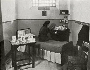
Background imageCell Collection: Girls room, Aylesbury Borstal, Buckinghamshire

Girls room, Aylesbury Borstal, Buckinghamshire
A typical inmates room at the Aylesbury girls borstal, Buckinghamshire, established in 1909 as a youth prison for those aged 16 to 21

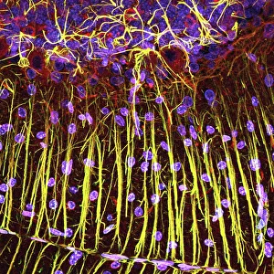
Background imageCell Collection: Cerebellum tissue, light micrograph

Cerebellum tissue, light micrograph
Cerebellum tissue. Confocal light micrograph of a section through the cerebellum of the brain. Purkinje cells, a type of neuron (nerve cell), are red

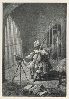
Background imageCell Collection: Man in the Iron Mask, playing the cello in prison

Man in the Iron Mask, playing the cello in prison
There are conflicting theories as to who this man was. One is that he was Count Ercole Antonio Mattioli (Matthioli or Marchioly, 1640-1703), an Italian diplomat, arrested as Eustache Dauger in 1669

Background imageCell Collection: Antibodies attacking virus particles, illustration

Antibodies attacking virus particles, illustration
3d illustration of antibodies attacking virus particles in the bloodstream

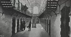
Background imageCell Collection: Portsmouth Prison Gallery

Portsmouth Prison Gallery
One of the galleries at Portsmouth Prison, Hampshire, cell doors lining each side. Date: circa 1900

Background imageCell Collection: Microscopic view of human respiratory syncytial virus

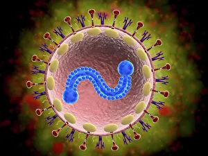
Microscopic view of human respiratory syncytial virus (RSV). RSV causes respiratory tract infection of the lungs and breathing passages

Background imageCell Collection: Activated macrophage, SEM C015 / 6375

Activated macrophage, SEM C015 / 6375
Activated macrophage. Coloured scanning electron micrograph (SEM) of an active macrophage white blood cell. Macrophages are cells of the bodys immune system that are found in the tissues rather than

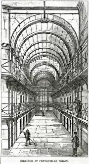
Background imageCell Collection: Cell wing at Pentonville Prison

Cell wing at Pentonville Prison. Date: 1862

Background imageCell Collection: IUD contraceptive and sperm cells

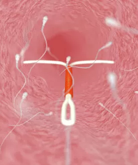
IUD contraceptive and sperm cells. Computer artwork of an intrauterine device (IUD, red and white, centre) in a womans uterus, surrounded by sperm cells (white, not to scale)

Background imageCell Collection: The Marquis de Sade (1740-1814) in Prison (engraving)

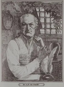
The Marquis de Sade (1740-1814) in Prison (engraving)
WYN138844 The Marquis de Sade (1740-1814) in Prison (engraving) by French School, (19th century); Private Collection; French, out of copyright

Background imageCell Collection: Anatomy, Biology, Blood Vessel, Cell, Color Image, Fibrin, Healthcare And Medicine

Anatomy, Biology, Blood Vessel, Cell, Color Image, Fibrin, Healthcare And Medicine
Anatomy, Biology, Blood Vessel, Cell, Color Image, Fibrin, Healthcare, Science Photo Library, 85758208

Background imageCell Collection: Picture No. 11675582

Picture No. 11675582
Scanning Electron micrograph (SEM): Lily Pollen grains. Date:

Background imageCell Collection: Picture No. 11675585

Picture No. 11675585
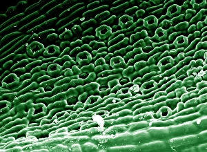
Scanning Electron micrograph (SEM)showing stomata on a Yew Leaf. Date:

Background imageCell Collection: Embryo development 24-36 hours after fertilization

Embryo development 24-36 hours after fertilization

Background imageCell Collection: Louis XVI (1754-93) Taking Leave of his Wife and Family, 1793 (coloured engraving)

Louis XVI (1754-93) Taking Leave of his Wife and Family, 1793 (coloured engraving)
XIR238623 Louis XVI (1754-93) Taking Leave of his Wife and Family, 1793 (coloured engraving) by Gillray, James (1757-1815); Musee de la Ville de Paris, Musee Carnavalet, Paris

Background imageCell Collection: Picture No. 10873704

Picture No. 10873704
Sleeping Sickness Parasite in red blood cells (Trypanosoma sp. ) Date:

Background imageCell Collection: Acute promyelocytic leukaemia, micrograph

Acute promyelocytic leukaemia, micrograph
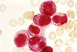
Acute promyelocytic leukaemia. Light micrograph of blood cells from bone marrow in a case of acute promyelocytic leukaemia. Leukaemia is a cancer where certain blood cells form in excess

Background imageCell Collection: Rod and cone cells of the eye, SEM C014 / 4866

Rod and cone cells of the eye, SEM C014 / 4866
Rod and cone cells of the eye. Coloured scanning electron micrograph (SEM) of rod and cone cells in the retina of a mammalian eye

Background imageCell Collection: Zinc fingers bound to a DNA strand

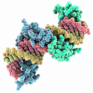
Zinc fingers bound to a DNA strand, molecular model. The double helix of DNA (deoxyribonucleic acid, red and yellow) is seen here with two Zif268 proteins (blue and green)

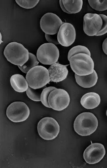
Background imageCell Collection: Human red blood corpuscles

Human red blood corpuscles
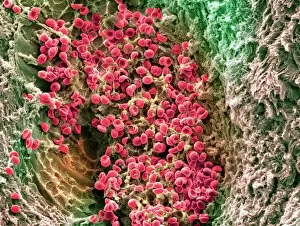
Scanning electron microscope (SEM) of red blood cells showing their characteristic biconcave shape which increases the surface area for diffusion

Background imageCell Collection: Dividing cells

Dividing cells. Computer artwork of a spiral of cells during mitosis (nuclear division). During mitosis two daughter nuclei are formed from one parent nucleus

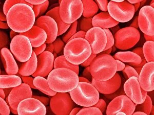
Background imageCell Collection: Red blood cells, SEM

Red blood cells, SEM
Red blood cells, coloured scanning electron micrograph (SEM). Red blood cells (erythrocytes) are biconcave, giving them a large surface area for gas exchange, and highly elastic

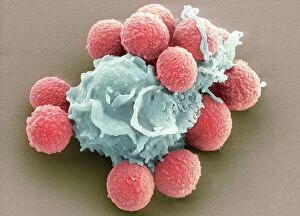
Background imageCell Collection: Phagocytosis of fungal spores, SEM

Phagocytosis of fungal spores, SEM
Phagocytosis of a fungus spore. Coloured scanning electron micrograph (SEM) of a neutrophil (blue) phagocytosing (engulfing and destroying) spores from the fungus Aspergillus fumigatus (red)

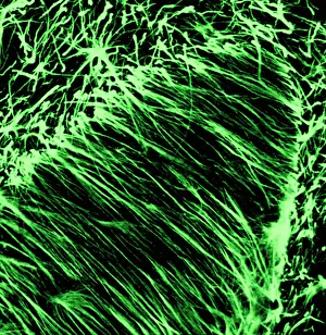
Background imageCell Collection: Oligodendrocyte nerve cells

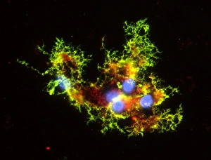
Oligodendrocyte nerve cells. Fluorescent light micrograph of human oligodendrocyte nerve cells. Cell nuclei, which contain the cells genetic information, have been dyed blue


All Professionally Made to Order for Quick Shipping

"Exploring the Intricacies of Life: Unveiling the Wonders of Cells" Delving into the microscopic world, a histological diagram of a mammalian retina reveals the intricate structure and organization of cells that enable us to perceive light. The cerebellum tissue, captured in a light micrograph, showcases its complex network of cells responsible for coordinating movement and balance. Intriguingly, even philosophers find solace in contemplating cellular existence. "Philosopher in Meditation, " an oil painting from 1632, reminds us that pondering life's mysteries often leads us back to our fundamental building blocks – cells. Zooming further into this realm, nerve and glial cells come alive under the lens. This captivating light micrograph highlights their vital role in transmitting signals throughout our nervous system. Meanwhile, a lavender pollen grain captured through scanning electron microscopy (SEM) displays nature's exquisite design at the cellular level. The battle against diseases takes center stage as T lymphocytes confront cancer cells in another SEM image. Science and evolution intertwine as we witness these tiny warriors fighting for survival within our bodies. Artistic expressions also shed light on cell significance; "The Death of Socrates, " painted in 1787, serves as a poignant reminder that even great minds are ultimately composed of countless individual cells. Santiago Ramon y Cajal's meticulous drawing from 1894 unveils various cell types within the mammalian cerebellum. His work not only represents scientific progress but also exemplifies how art can aid understanding by visually capturing complexity. Stepping away from biology momentarily, we encounter Rolls Royce/Snecma Olympus 593 Mk602 engine undergoing testing within a controlled environment called a test cell. Here too, precision engineering relies on understanding cellular mechanics to achieve optimal performance. Returning to biological marvels - HeLa cells take center stage under yet another microscope lens. These immortalized human cells have revolutionized medical research, paving the way for countless breakthroughs.