Fraud Blocker Skip to main content

Cell Collection (page 7)

"Exploring the Intricacies of Life: Unveiling the Wonders of Cells" Delving into the microscopic world

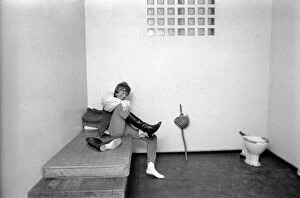
Background imageCell Collection: Tommy Steele who rode on the top of a stage coach as Dick Whittington in the Lord Mayors

Tommy Steele who rode on the top of a stage coach as Dick Whittington in the Lord Mayors show changed, after travelling through City of London in the procession

Background imageCell Collection: Convair F-106B-31-CO Delta Dart 57-2507

Convair F-106B-31-CO Delta Dart 57-2507
Convair NF-106B-31-CO Delta Dart 57-2507 (msn 8-27-01) at NASA Lewis Research Center, Lewis Field, Cleveland, OH. registered as NASA 607 Built April 1958

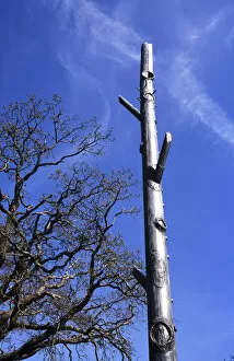
Background imageCell Collection: Communications, Telephone, Mast

Communications, Telephone, Mast
Communications, Telephone, Mobile phone mast disguised to look like a tree, Tunbridge Wells, Kent, England

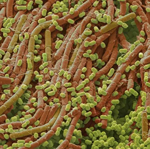
Background imageCell Collection: Soil bacteria, SEM

Soil bacteria, SEM
Soil bacteria. Coloured scanning electron micrograph (SEM). Bacteria in the soil are directly tied to nutrient recycling especially carbon, nitrogen, phosphorus and sulfur

Background imageCell Collection: Hollyhock pollen grain, SEM

Hollyhock pollen grain, SEM
Hollyhock pollen grain. Coloured scanning electron micrograph (SEM) of a pollen grain from a hibiscus (Alcea setosa) flower. Pollen grains are the male sex cells of a flowering plant

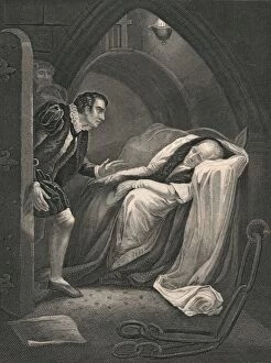
Background imageCell Collection: The Death of Mortimer. (mid 19th century). Creator: J Rogers

The Death of Mortimer. (mid 19th century). Creator: J Rogers
The Death of Mortimer. (mid 19th century). Scene from Shakespeares " Henry VI", (act II scene V). Richard Plantagenet enters a cell in the Tower of London to find Edmund Mortimer lying

Background imageCell Collection: Napoleon in prison at Nice, France, 1794. Artist: J Outrim

Napoleon in prison at Nice, France, 1794. Artist: J Outrim
Napoleon in prison at Nice, France, 1794. Napoleon Bonaparte (1721-1818) enjoyed a meteoric rise through the ranks of the French Revolutionary army

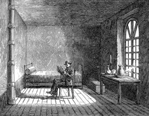
Background imageCell Collection: Paris Demolitions - prisoner's chamber, 1864. Creator: Unknown

Paris Demolitions - prisoner's chamber, 1864. Creator: Unknown
Paris Demolitions - the Hotel des Haricots, or prison of the National Guard of Paris: prisoner's chamber, 1864. Interior of '...the Hotel des Haricots

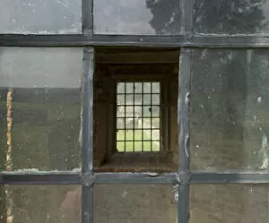
Background imageCell Collection: Window detail DP168288

Window detail DP168288
Hamsterley Hall, Hamsterley Mill, Rowlands Gill, County Durham. Detail looking through a window in the summerhouse, formerly a cupola from Beaudesert, Staffordshire, in the houses east garden

Background imageCell Collection: South East Asia, Thailand, Buddhist monk using a lap top and a mobile phone

South East Asia, Thailand, Buddhist monk using a lap top and a mobile phone

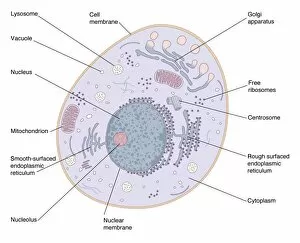
Background imageCell Collection: Animal cell structure

Animal cell structure. Artwork of a section through an animal cell. At lower centre is the nucleus, which contains the cells genetic information in the form of DNA (deoxyribonucleic acid)

Background imageCell Collection: Personal Jesus

Personal Jesus
Petri Damstén

Background imageCell Collection: Preserved Heart

Preserved Heart
Petri Damstén

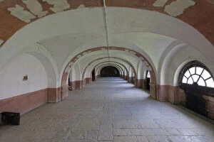
Background imageCell Collection: Prison cells, Clairvaux Abbey, Ville-sous-la-Ferte, Aube

Prison cells, Clairvaux Abbey, Ville-sous-la-Ferte, Aube
Group cells in the men's prison, (the abbey functioned as a prison from 1804 when it was converted by Napoleon), at the Abbaye de Clairvaux, or Clairvaux Abbey

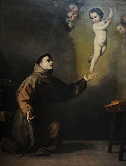
Background imageCell Collection: Saint Anthony of Padua (1195-1231) by Juseppe de Ribera

Saint Anthony of Padua (1195-1231) by Juseppe de Ribera
Saint Anthony of Padua (1195-1231). Portuguese friar of Franciscan Order. Painting by Juseppe de Ribera (1591-1652). Spanish painter. San Fernando Royal Academy of Fine Arts. Madrid. Spain. Date: 2019
Background imageCell Collection: Napoleon in the Prison of Nice

Napoleon in the Prison of Nice, France. Date: 1794

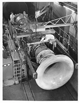
Background imageCell Collection: Rolls-Royce Spey turbofan engine

Rolls-Royce Spey turbofan engine in a test cell fitted with an experimental afterburner, on 1 April 1963. Date: 1962

Background imageCell Collection: Suffragette in Prison Cell

Suffragette in Prison Cell. Cover illustration for Die Suffragettes by Kaethe Schirmacher (Alexander Duncker Verlag Weimar 1912) Based on a sketch made in Holloway Prison by Sylvia Pankhurst

Background imageCell Collection: These are scenes from the London Orphan Asylum in Watford. 1) boys doing drill 2) the boys band 3

These are scenes from the London Orphan Asylum in Watford. 1) boys doing drill 2) the boys band 3) sewing class 4) the Prince of Wales proposing the toast of the asylum at the Jubilee Dinner at

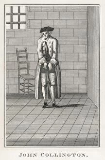
Background imageCell Collection: JOHN COLLINGTON

JOHN COLLINGTON: Ne-er-do-well & wife beater, poacher, hiring hitmen & getting others to menace his neighbours. Buried 6 of his dead children in the orchard to save money! Date: 1696/7 - 1750

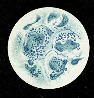
Background imageCell Collection: Colony of Haemophilus influenzae, 1906 (litho)

Colony of Haemophilus influenzae, 1906 (litho)
3501914 Colony of Haemophilus influenzae, 1906 (litho) by German School, (20th century); Private Collection; (add.info.: Bacteria constitute a large domain of prokaryotic microorganisms)

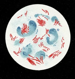
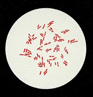
Background imageCell Collection: Colony of Mycobacterium leprae, 1906 (litho)

Colony of Mycobacterium leprae, 1906 (litho)
3501912 Colony of Mycobacterium leprae, 1906 (litho) by German School, (20th century); Private Collection; (add.info.: Bacteria constitute a large domain of prokaryotic microorganisms)

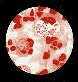
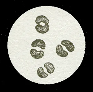
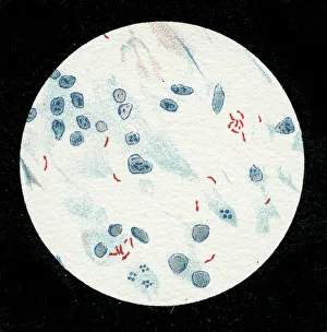
Background imageCell Collection: Colony of Micrococcus Gonorrhoea, 1906 (litho)

Colony of Micrococcus Gonorrhoea, 1906 (litho)
3501903 Colony of Micrococcus Gonorrhoea, 1906 (litho) by German School, (20th century); Private Collection; (add.info.: Bacteria constitute a large domain of prokaryotic microorganisms)

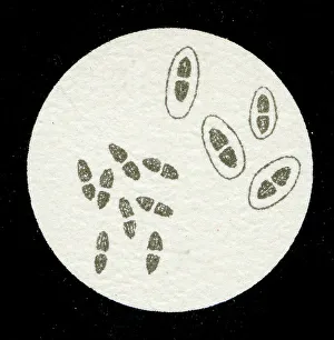
Background imageCell Collection: Colony of Streptococcus pneumoniae with and without bubble capsule, 1906 (litho)

Colony of Streptococcus pneumoniae with and without bubble capsule, 1906 (litho)
3501899 Colony of Streptococcus pneumoniae with and without bubble capsule, 1906 (litho) by German School, (20th century); Private Collection; (add.info)

Background imageCell Collection: Colony of Streptococcus Pneumoniae, 1906 (litho)

Colony of Streptococcus Pneumoniae, 1906 (litho)
3501898 Colony of Streptococcus Pneumoniae, 1906 (litho) by German School, (20th century); Private Collection; (add.info.: Bacteria constitute a large domain of prokaryotic microorganisms)

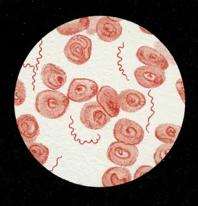
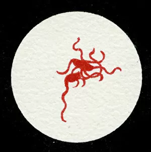
Background imageCell Collection: Spirillum of the Lyme disease causing bacteria, Spirochaetes Borrelia Recurrentis

Spirillum of the Lyme disease causing bacteria, Spirochaetes Borrelia Recurrentis, in blood sample, 1906 (litho)
3501919 Spirillum of the Lyme disease causing bacteria, Spirochaetes Borrelia Recurrentis, in blood sample, 1906 (litho) by German School

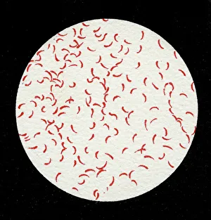
Background imageCell Collection: Colony of Vibrio cholerae, 1906 (litho)

Colony of Vibrio cholerae, 1906 (litho)
3501918 Colony of Vibrio cholerae, 1906 (litho) by German School, (20th century); Private Collection; (add.info.: Bacteria constitute a large domain of prokaryotic microorganisms)

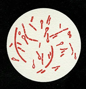
Background imageCell Collection: Colony of Clostridium tetani with spores, 1906 (litho)

Colony of Clostridium tetani with spores, 1906 (litho)
3501913 Colony of Clostridium tetani with spores, 1906 (litho) by German School, (20th century); Private Collection; (add.info.: Bacteria constitute a large domain of prokaryotic microorganisms)

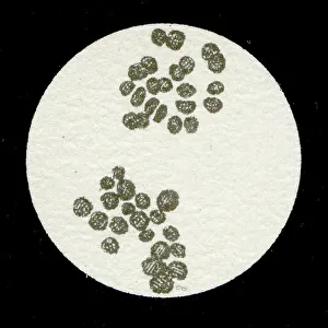
Background imageCell Collection: Colony of Mycobacterium tuberculosis, 1906 (litho)

Colony of Mycobacterium tuberculosis, 1906 (litho)
3501910 Colony of Mycobacterium tuberculosis, 1906 (litho) by German School, (20th century); Private Collection; (add.info.: Bacteria constitute a large domain of prokaryotic microorganisms)

Background imageCell Collection: A colony of Staphylococcus pyogenes after cell division, 1906 (litho)

A colony of Staphylococcus pyogenes after cell division, 1906 (litho)
3501897 A colony of Staphylococcus pyogenes after cell division, 1906 (litho) by German School, (20th century); Private Collection; (add.info)

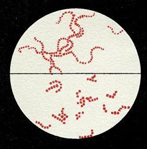
Background imageCell Collection: Colony of Streptococcus pyogenes, 1906 (litho)

Colony of Streptococcus pyogenes, 1906 (litho)
3501894 Colony of Streptococcus pyogenes, 1906 (litho) by German School, (20th century); Private Collection; (add.info.: Bacteria constitute a large domain of prokaryotic microorganisms)

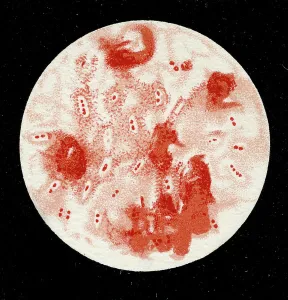
Background imageCell Collection: Colony of Yersinia pestis, 1906 (litho)

Colony of Yersinia pestis, 1906 (litho)
3501917 Colony of Yersinia pestis, 1906 (litho) by German School, (20th century); Private Collection; (add.info.: Bacteria constitute a large domain of prokaryotic microorganisms)

Background imageCell Collection: Colony of Salmonella Typhi showing Bacilli with flagella, 1906 (litho)

Colony of Salmonella Typhi showing Bacilli with flagella, 1906 (litho)
3501916 Colony of Salmonella Typhi showing Bacilli with flagella, 1906 (litho) by German School, (20th century); Private Collection; (add.info)

Background imageCell Collection: Colony of Salmonella typhi, 1906 (litho)

Colony of Salmonella typhi, 1906 (litho)
3501915 Colony of Salmonella typhi, 1906 (litho) by German School, (20th century); Private Collection; (add.info.: Bacteria constitute a large domain of prokaryotic microorganisms)

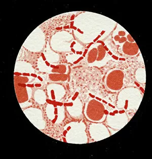
Background imageCell Collection: Colony of Bacillus Anthracis in a human spleen, 1906 (litho)

Colony of Bacillus Anthracis in a human spleen, 1906 (litho)
3501905 Colony of Bacillus Anthracis in a human spleen, 1906 (litho) by German School, (20th century); Private Collection; (add.info)

Background imageCell Collection: Spores of Bacillus Anthracis, 1906 (litho)

Spores of Bacillus Anthracis, 1906 (litho)
3501904 Spores of Bacillus Anthracis, 1906 (litho) by German School, (20th century); Private Collection; (add.info.: Bacteria constitute a large domain of prokaryotic microorganisms)

Background imageCell Collection: Colony of Micrococcus Gonorrhoea found in the Urethra, 1906 (litho)

Colony of Micrococcus Gonorrhoea found in the Urethra, 1906 (litho)
3501900 Colony of Micrococcus Gonorrhoea found in the Urethra, 1906 (litho) by German School, (20th century); Private Collection; (add.info)

Background imageCell Collection: Colony of Corynebacterium Diphtheriae, 1906 (litho)

Colony of Corynebacterium Diphtheriae, 1906 (litho)
3501907 Colony of Corynebacterium Diphtheriae, 1906 (litho) by German School, (20th century); Private Collection; (add.info.: Bacteria constitute a large domain of prokaryotic microorganisms)

Background imageCell Collection: Colony of Bacterium mallei, 1906 (litho)

Colony of Bacterium mallei, 1906 (litho)
3501906 Colony of Bacterium mallei, 1906 (litho) by German School, (20th century); Private Collection; (add.info.: Bacteria constitute a large domain of prokaryotic microorganisms)

Background imageCell Collection: Staphylococcus colony, 1905 (litho)

Staphylococcus colony, 1905 (litho)
3501895 Staphylococcus colony, 1905 (litho) by German School, (20th century); Private Collection; (add.info.: Bacteria constitute a large domain of prokaryotic microorganisms)

Background imageCell Collection: Colony of Mycobacterium Tuberculosis, 1906 (litho)

Colony of Mycobacterium Tuberculosis, 1906 (litho)
3501911 Colony of Mycobacterium Tuberculosis, 1906 (litho) by German School, (20th century); Private Collection; (add.info.: Bacteria constitute a large domain of prokaryotic microorganisms)

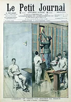
Background imageCell Collection: Cell and Library at the prison at Fresnes, 1907 (print)

Cell and Library at the prison at Fresnes, 1907 (print)
537582 Cell and Library at the prison at Fresnes, 1907 (print) by French School, (20th century); (add.info.: From Le Petit Journal, Paris, 9 November 1907)

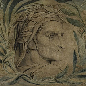
Background imageCell Collection: Dante Alighieri, c. 1800-03 (pen & ink with tempera on canvas)

Dante Alighieri, c. 1800-03 (pen & ink with tempera on canvas)
1113188 Dante Alighieri, c.1800-03 (pen & ink with tempera on canvas) by Blake, William (1757-1827); Manchester Art Gallery

Background imageCell Collection: Dante Alighieri, c. 1800-03 (pen & ink with tempera on canvas)

Dante Alighieri, c. 1800-03 (pen & ink with tempera on canvas)
MAN350937 Dante Alighieri, c.1800-03 (pen & ink with tempera on canvas) by Blake, William (1757-1827); 42.5x87.8 cm; Manchester Art Gallery

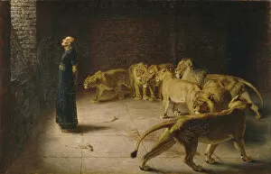
Background imageCell Collection: Daniel's Answer to the King, 1890 (oil on canvas)

Daniel's Answer to the King, 1890 (oil on canvas)
3082374 Daniel's Answer to the King, 1890 (oil on canvas) by Riviere, Briton (1840-1920); 120.5x187.9 cm; Manchester Art Gallery

Background imageCell Collection: Miss Lucy Burns in Occoquan Workhouse, Washington, 1917 (b/w photo)

Miss Lucy Burns in Occoquan Workhouse, Washington, 1917 (b/w photo)
746438 Miss Lucy Burns in Occoquan Workhouse, Washington, 1917 (b/w photo) by Harris & Ewing (1905-45); Private Collection; (add.info)

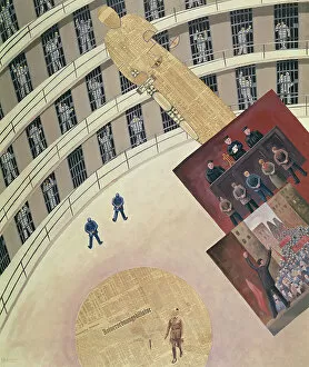
Background imageCell Collection: The Political Prisoners, 1931 (collage)

The Political Prisoners, 1931 (collage)
ALG158881 The Political Prisoners, 1931 (collage) by Fuhrmann, Paul (1893-1952); Museum fur Deutsche Geschichte, Berlin, Germany; (add.info.: artist imprisoned for anti-militaristic views;)

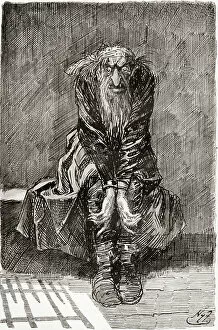
Background imageCell Collection: Fagin in the Condemned Cell. ' He sat down on a stone bench opposite the door

Fagin in the Condemned Cell. " He sat down on a stone bench opposite the door
1000821 Fagin in the Condemned Cell. " He sat down on a stone bench opposite the door, which served for a seat and bedstead, and casting his bloodshot eyes upon the grond


All Professionally Made to Order for Quick Shipping

"Exploring the Intricacies of Life: Unveiling the Wonders of Cells" Delving into the microscopic world, a histological diagram of a mammalian retina reveals the intricate structure and organization of cells that enable us to perceive light. The cerebellum tissue, captured in a light micrograph, showcases its complex network of cells responsible for coordinating movement and balance. Intriguingly, even philosophers find solace in contemplating cellular existence. "Philosopher in Meditation, " an oil painting from 1632, reminds us that pondering life's mysteries often leads us back to our fundamental building blocks – cells. Zooming further into this realm, nerve and glial cells come alive under the lens. This captivating light micrograph highlights their vital role in transmitting signals throughout our nervous system. Meanwhile, a lavender pollen grain captured through scanning electron microscopy (SEM) displays nature's exquisite design at the cellular level. The battle against diseases takes center stage as T lymphocytes confront cancer cells in another SEM image. Science and evolution intertwine as we witness these tiny warriors fighting for survival within our bodies. Artistic expressions also shed light on cell significance; "The Death of Socrates, " painted in 1787, serves as a poignant reminder that even great minds are ultimately composed of countless individual cells. Santiago Ramon y Cajal's meticulous drawing from 1894 unveils various cell types within the mammalian cerebellum. His work not only represents scientific progress but also exemplifies how art can aid understanding by visually capturing complexity. Stepping away from biology momentarily, we encounter Rolls Royce/Snecma Olympus 593 Mk602 engine undergoing testing within a controlled environment called a test cell. Here too, precision engineering relies on understanding cellular mechanics to achieve optimal performance. Returning to biological marvels - HeLa cells take center stage under yet another microscope lens. These immortalized human cells have revolutionized medical research, paving the way for countless breakthroughs.