
Inhibition Collection
"Inhibition: Unveiling the Complexities of Control" Inhibition, a multifaceted concept, encompasses various realms of human existence


All Professionally Made to Order for Quick Shipping
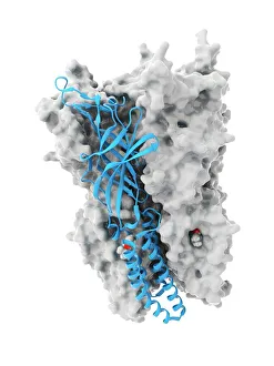
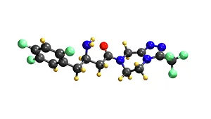
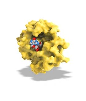
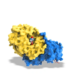
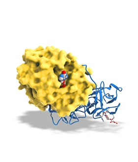
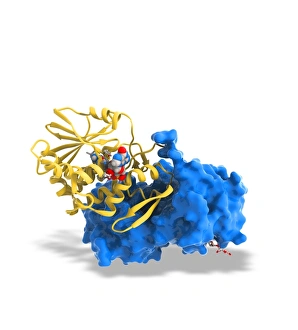
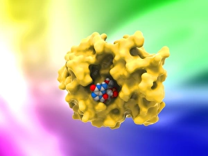
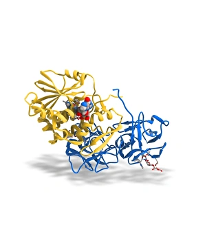
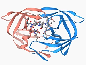
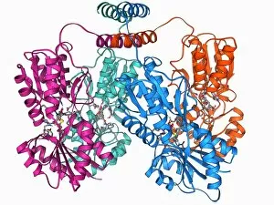
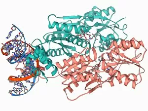
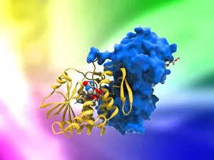
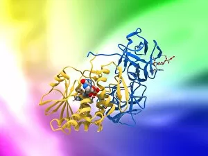
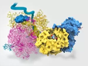
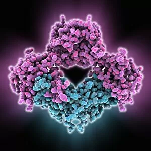
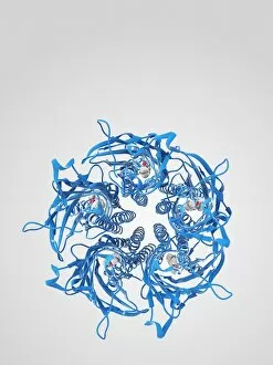
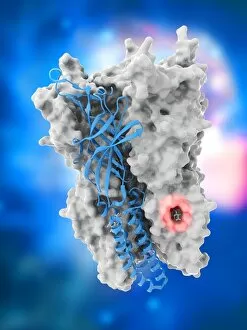
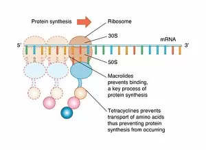
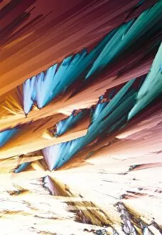
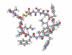
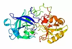
"Inhibition: Unveiling the Complexities of Control" Inhibition, a multifaceted concept, encompasses various realms of human existence. From the intricate workings of our bodies to artistic expressions and societal dynamics, inhibition shapes our experiences in profound ways. At the molecular level, we witness inhibition's power through anaesthetics that target ion channels like C015/6718. These remarkable substances selectively block these channels, temporarily numbing pain and allowing medical procedures to be performed with ease. Similarly, Januvia diabetes drug molecule stands as a testament to inhibition's potential for improving lives. By inhibiting specific enzymes involved in glucose regulation, this medication helps manage blood sugar levels for individuals battling diabetes. Beyond science and medicine lies the realm of artistry. "The Jester; Le Bouffon" by an unknown artist from 1868 captures the essence within its strokes on canvas. The jester's playful facade conceals layers of restraint and self-censorship necessary to navigate social expectations during that era. Moving back into scientific territory, we encounter Ricin A-chain (artwork C017/3653) alongside several depictions (C017/3652-3651). These artworks unveil how ricin molecules inhibit protein synthesis within cells—a chilling reminder of nature's potent mechanisms capable of causing harm if not understood or controlled. However, not all forms are detrimental. In fact, some conceptualize it as a loss—depicted eloquently in artwork F007/8262 titled "Loss of Inhibition. " This thought-provoking piece invites contemplation about breaking free from societal constraints while acknowledging potential consequences when boundaries dissolve entirely. Returning once more to ricin-related artworks (C017/3654-3649), we delve deeper into understanding its structure and function—an essential step towards developing countermeasures against this deadly toxin.