
Tumour Suppressor Protein Collection
The tumour suppressor protein, also known as p53, plays a crucial role in preventing the development and progression of cancer


All Professionally Made to Order for Quick Shipping
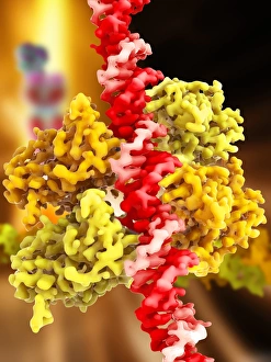
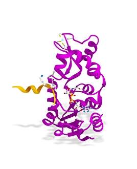
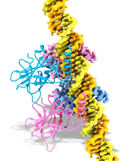
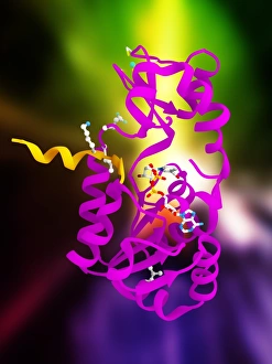
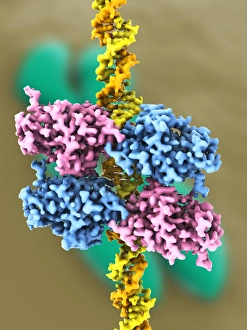
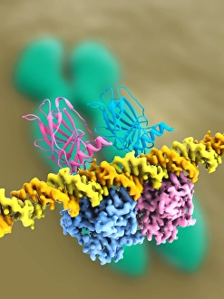
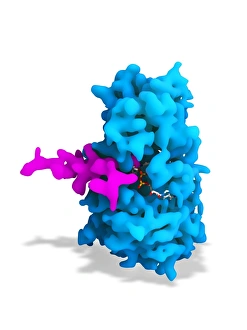
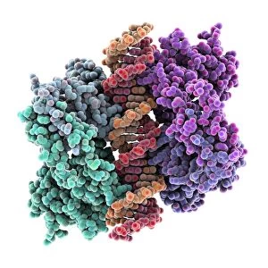
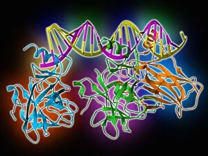
The tumour suppressor protein, also known as p53, plays a crucial role in preventing the development and progression of cancer. It acts as a guardian of the genome by monitoring DNA integrity and initiating repair mechanisms when necessary. The intricate relationship between this protein and DNA is depicted in captivating artwork C017 / 3647, where they intertwine to symbolize their close connection. Another key player in this process is the Sirtuin enzyme, which collaborates with p53 to regulate gene expression and maintain genomic stability. This partnership is beautifully portrayed in artwork C017 / 3659, showcasing their synchronized dance within the cellular environment. Intriguingly, further illustrations such as C017 / 3644 and C017 / 3658 depict different perspectives of the interaction between p53 and DNA. These visuals highlight the complexity of this relationship while emphasizing its significance for tumor suppression. Delving deeper into molecular structures, images like C017 / 3646, C017 / 3645, F006 / 9729, F006 /9564, and F006/9523 provide detailed representations of both p53 and DNA complexes. These models help scientists understand how mutations or alterations can disrupt normal functioning and lead to uncontrolled cell growth characteristic of cancer. Lastly, F006/9451 presents an intriguing molecular model that showcases various components involved in tumour suppression. This visual aids researchers in comprehending the intricate mechanisms underlying this vital biological process.