Jigsaw Puzzle : Fungal reproduction, SEM


![]()
Jigsaw Puzzles from Science Photo Library
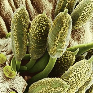
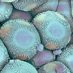
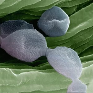
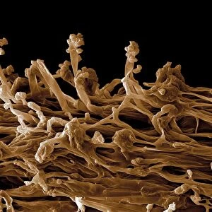
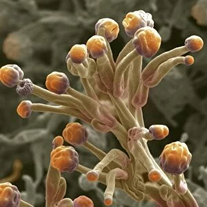
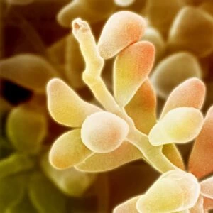
Fungal reproduction, SEM
Fungal reproduction. Coloured scanning electron micrograph (SEM) of the tip of a branch of a conidiophore (one type of fungal reproductive structure) with a conidia (spore) emerging from the tip. The spore is a reproductive structure that is released to drift in the air and land elsewhere to form a new fungal body. This is a conidiophore from the pathogenic fungus Fusarium oxysporum, which causes wilt disease in tomato and carnation plants
Science Photo Library features Science and Medical images including photos and illustrations
Media ID 6292241
© DR JEREMY BURGESS/SCIENCE PHOTO LIBRARY
Conidia Conidiophore Eumycota Fungal Fungi Fungus Mycology Re Production Reproducing Reproductive Spore False Coloured Micro Biology Microbiological Pathogen
Jigsaw Puzzle (500 Pieces)
Discover the intricacies of the natural world with our Media Storehouse Jigsaw Puzzles. This scientific puzzle, featuring a captivating Coloured Scanning Electron Micrograph (SEM) image from Science Photo Library, invites you to explore the fascinating world of Fungal Reproduction. Delve into the details of this conidiophore branch, observing the emergence of a conidium (spore) from its tip. A stimulating and educational pastime for puzzle enthusiasts and biology aficionados alike.
500 piece puzzles are custom made in Australia and hand-finished on 100% recycled 1.6mm thick laminated puzzle boards. There is a level of repetition in jigsaw shapes with each matching piece away from its pair. The completed puzzle measures 40x51cm and is delivered packaged in an attractive presentation box specially designed to fit most mail slots with a unique magnetic lid
Jigsaw Puzzles are an ideal gift for any occasion
Estimated Product Size is 50.7cm x 40.3cm (20" x 15.9")
These are individually made so all sizes are approximate
Artwork printed orientated as per the preview above, with landscape (horizontal) or portrait (vertical) orientation to match the source image.
EDITORS COMMENTS
This print showcases the intricate process of fungal reproduction. In this coloured scanning electron micrograph (SEM), we are granted a close-up view of a conidiophore, one type of fungal reproductive structure. At the tip of a branch, we witness the emergence of a conidia, which is essentially a spore responsible for generating new fungal bodies. The significance lies in understanding that these spores are released into the air and eventually settle elsewhere to establish fresh fungal entities. The image captures this delicate moment when life takes flight and disperses to create anew. Specifically, this conidiophore belongs to Fusarium oxysporum, an infamous pathogenic fungus notorious for causing wilt disease in tomato and carnation plants. Its ability to reproduce through these structures contributes greatly to its destructive nature. Through false-coloured enhancement techniques employed by scanning electron microscopy (SEM), we can appreciate the beauty hidden within biology's microscopic realm. This photograph not only highlights the complexity and diversity found within fungi but also serves as a reminder of their crucial role in our ecosystem. Science Photo Library has once again provided us with an awe-inspiring glimpse into nature's wonders, allowing us to marvel at its intricacies and expand our knowledge in fields such as microbiology and mycology.
MADE IN AUSTRALIA
Safe Shipping with 30 Day Money Back Guarantee
FREE PERSONALISATION*
We are proud to offer a range of customisation features including Personalised Captions, Color Filters and Picture Zoom Tools
SECURE PAYMENTS
We happily accept a wide range of payment options so you can pay for the things you need in the way that is most convenient for you
* Options may vary by product and licensing agreement. Zoomed Pictures can be adjusted in the Cart.