
Conidiophore Collection
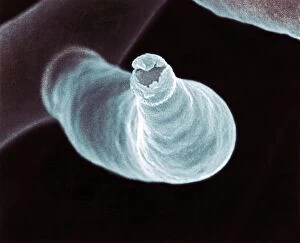
"Exploring the Intricate World of Conidiophore: A Close-up Look at Fungal Structures" In this captivating image (Picture No

All Professionally Made to Order for Quick Shipping
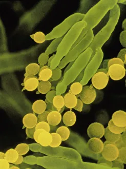
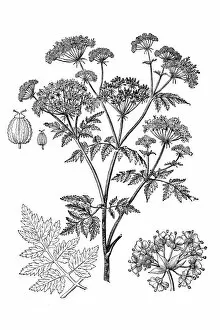
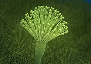
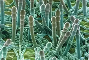
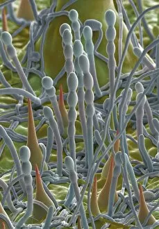
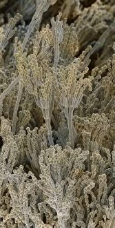
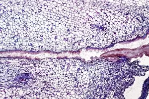
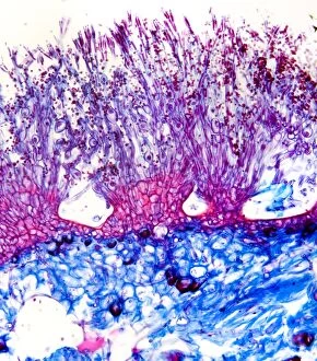
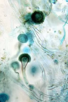
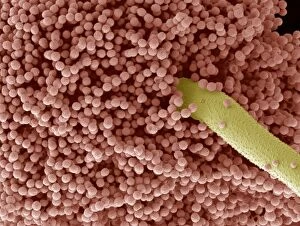
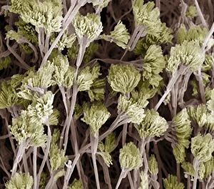
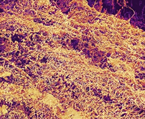
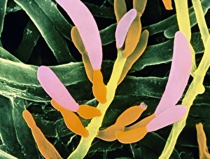
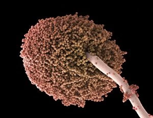
"Exploring the Intricate World of Conidiophore: A Close-up Look at Fungal Structures" In this captivating image (Picture No. 11014633), captured using a scanning electron microscope (SEM), we delve into the fascinating realm of conidiophores, specifically those found in the penicillin fungus. These slender structures serve as reproductive organs, producing and dispersing countless spores that aid in fungal propagation. Conium maculatum, commonly known as hemlock or poison hemlock, is another plant species associated with conidiophores. Its intricate network of these specialized structures can be observed under SEM, revealing their role in spreading toxic spores. Artwork C013 / 4613 showcases an Aspergillus fungus's conidiophore arrangement. This artwork beautifully illustrates how these elongated stalks bear clusters of tiny spore-producing cells called conidia at their tips. Powdery mildew on a leaf is a common sight in gardens and crops worldwide. SEM imagery provides us with an up-close view of powdery mildew's unique conidiophores and the powdery appearance they create on infected leaves. Penicillium fungal spores are renowned for their medicinal properties. SEM images allow us to appreciate the delicate structure of penicillium's conidiophores responsible for generating these valuable therapeutic agents. Grey mould fungus also exhibits its own distinct array of branching conidiophores when viewed under light microscopy. Multiple light micrographs showcase this mold's ability to produce abundant spore-bearing structures essential for its survival and spread. Similarly, brown mould fungus displays intriguing patterns through light microscopy examination—concerning yet mesmerizing formations that highlight nature's diversity within fungal life cycles. As we explore various microscopic perspectives on different fungi species' conidiophores, we gain insight into their vital roles in reproduction and dispersion strategies across diverse ecosystems.