Photo Mug : Pancreas cells, SEM


![]()
Home Decor from Science Photo Library
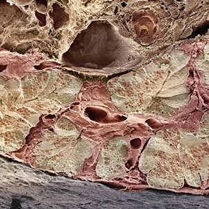
Pancreas cells, SEM
Pancreas cells. Coloured scanning electron micrograph (SEM) of acinar (exocrine) pancreatic cells. Acinar cells produce and excrete digestive enzymes to the small intestine, via the pancreatic ducts. The enzymes are excreted in vesicles known as zymogen granules (red). The enzymes are activated in the small intestine and aid the breakdown of carbohydrates, fats and proteins. Magnification: x330 when printed 10 centimetres wide
Science Photo Library features Science and Medical images including photos and illustrations
Media ID 6450003
© STEVE GSCHMEISSNER/SCIENCE PHOTO LIBRARY
Digestive Enzymatic Enzyme Enzymes Excretory Exocrine False Colour Fracture Fractured Granules Histological Histology Pancreas Pancreatic Physiological Physiology Storage Transport Vesicle Vesicles Zymogen Granule False Coloured Section Sectioned
Photo Mug
Bring the wonders of science into your daily routine with Media Storehouse's Photo Mugs. Featuring an captivating image of Pancreas cells from Science Photo Library, this mug showcases the intricate details of acinar cells in a coloured scanning electron micrograph (SEM). These cells, essential for digestion, produce and excrete digestive enzymes to the small intestine. Each sip from this mug is a reminder of the marvels of the human body. Perfect for scientists, students, or anyone with a curious mind, this Photo Mug is a unique and thoughtful gift. Embrace the beauty of science, one mug at a time.
A personalised photo mug blends sentimentality with functionality, making an ideal gift for cherished loved ones, close friends, or valued colleagues. Preview may show both sides of the same mug.
Elevate your coffee or tea experience with our premium white ceramic mug. Its wide, comfortable handle makes drinking easy, and you can rely on it to be both microwave and dishwasher safe. Sold in single units, preview may show both sides of the same mug so you can see how the picture wraps around.
Mug Size is 8.1cm high x 9.6cm diameter (3.2" x 3.8")
These are individually made so all sizes are approximate
EDITORS COMMENTS
This print showcases the intricate beauty of pancreas cells, captured through a scanning electron microscope (SEM). The image is colored to highlight the various components and structures within these exocrine pancreatic cells. Acinar cells, depicted here in stunning detail, play a crucial role in our digestive system by producing and releasing digestive enzymes into the small intestine via specialized ducts. The zymogen granules, seen as vibrant red vesicles within the image, contain these powerful enzymes that aid in breaking down carbohydrates, fats, and proteins during digestion. Once activated in the small intestine, they contribute to our body's physiological processes. With a magnification of x330 when printed at 10 centimeters wide, this photograph allows us to appreciate the complexity and organization of these vital cellular structures. It offers an intriguing glimpse into the microscopic world that underlies our biological functions. Whether you are studying biology or simply fascinated by human anatomy and physiology, this visually striking SEM image provides both scientific insight and aesthetic appeal. Its false-colored presentation adds an artistic touch while maintaining its scientific integrity. This print from Science Photo Library serves as a reminder of how even at such minuscule scales, nature's wonders never cease to amaze us.
MADE IN AUSTRALIA
Safe Shipping with 30 Day Money Back Guarantee
FREE PERSONALISATION*
We are proud to offer a range of customisation features including Personalised Captions, Color Filters and Picture Zoom Tools
SECURE PAYMENTS
We happily accept a wide range of payment options so you can pay for the things you need in the way that is most convenient for you
* Options may vary by product and licensing agreement. Zoomed Pictures can be adjusted in the Cart.