Poster Print : Brain tumour, DTI MRI scan C017 / 7059

![]()
Poster Prints from Science Photo Library
Brain tumour, DTI MRI scan C017 / 7059
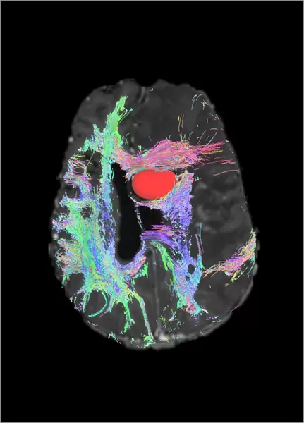
Brain tumour. Axial diffusion tensor imaging (DTI) magnetic resonance imaging (MRI) scan of nerve pathways (coloured) in a brain with a tumour (red, upper centre). The brain is seen from below, with the front of the brain at top. Brain tumours can be benign or malignant (cancerous). They can cause seizures, headaches, and memory and personality changes due to the growth of the tumour. The nerve fibres include the corticospinal tract (blue), passing from the motor cortex to the spinal cord. Diffusion tensor imaging (tractography) measures the direction of water diffusion, which in the brain reveals the orientation of nerve fibres
Science Photo Library features Science and Medical images including photos and illustrations
Media ID 9263955
© SHERBROOKE CONNECTIVITY IMAGING LAB/SCIENCE PHOTO LIBRARY
Brain Imaging Brain Scan Cancer Cancerous Central Nervous System Cerebral Diffusion Tensor Imaging Dti Scan Fiber Fibers Fibre Fibres Imaging Technique Magnetic Resonance Imaging Malignancy Malignant Motor Cortex Pathway Mri Scan Mri Scanner Nerve Nerve Fibre Nerves Neural Pathway Neural Tract Neuropathology Oncology Paths Pathway Pathways Structural Tractogram Tractography Tumor Tumour Brain Condition Disorder Neurological Neurology
A3 (42 x 29.7cm) Poster Print
Discover the intricacies of the human brain with our captivating selection of scientific poster prints from Media Storehouse. This particular print features an Axial Diffusion Tensor Imaging (DTI) Magnetic Resonance Imaging (MRI) scan of nerve pathways in a brain with a tumour, captured by SHERBROOKE CONNECTIVITY IMAGING LAB/SCIENCE PHOTO LIBRARY. The vibrant colours highlight the complex network of neural connections, contrasting the red tumour at the upper centre. Ignite curiosity and inspire learning in any space with this stunning, high-definition representation of brain anatomy.
Premium quality poster prints are printed on luxurious semi-gloss satin 270 gsm paper. Our meticulously crafted poster prints offer an affordable option for decorating any space, making them ideal for living rooms, bedrooms, offices and beyond. To ensure your poster arrives in good condition, we roll and send them in strong mailing tubes.
Poster prints are budget friendly enlarged prints in standard poster paper sizes (A0, A1, A2, A3 etc). Whilst poster paper is sometimes thinner and less durable than our other paper types, they are still ok for framing and should last many years. Our Archival Quality Photo Prints and Fine Art Paper Prints are printed on higher quality paper and the choice of which largely depends on your budget.
Estimated Product Size is 30.6cm x 42.6cm (12" x 16.8")
These are individually made so all sizes are approximate
Artwork printed orientated as per the preview above, with portrait (vertical) orientation to match the source image.
EDITORS COMMENTS
This print showcases a DTI MRI scan of a brain with a prominent red tumor at the upper center, against a striking black background. The image provides an intricate view of nerve pathways in vibrant colors, revealing the complex structure and disorder caused by the presence of this malignant growth. From below, we observe the brain with its front positioned at the top. This particular brain tumor can be either benign or cancerous (malignant), leading to various symptoms such as seizures, headaches, memory loss, and personality changes due to its expansion. The colored nerve fibers depicted include the corticospinal tract in blue, which connects the motor cortex to the spinal cord. Through diffusion tensor imaging (DTI) or tractography technique employed here, water diffusion direction is measured within the brain to unveil nerve fiber orientation. This image serves as an invaluable tool for medical professionals specializing in neurology and oncology. It offers crucial insights into neuropathology and aids in diagnosing and understanding conditions related to neural tracts. By visualizing these intricate pathways affected by tumors like this one through advanced MRI scanning techniques such as DTI scan or diffusion tensor imaging, healthcare practitioners gain valuable information necessary for treatment planning and patient care. Courtesy of Sherbrooke Connectivity Imaging Lab/Science Photo Library's extensive collection on medical imagery; this photograph stands testament to their commitment towards advancing scientific knowledge about neurological disorders while showcasing breathtaking beauty hidden within our own bodies.
MADE IN AUSTRALIA
Safe Shipping with 30 Day Money Back Guarantee
FREE PERSONALISATION*
We are proud to offer a range of customisation features including Personalised Captions, Color Filters and Picture Zoom Tools
SECURE PAYMENTS
We happily accept a wide range of payment options so you can pay for the things you need in the way that is most convenient for you
* Options may vary by product and licensing agreement. Zoomed Pictures can be adjusted in the Cart.