Home > Science > Xray
Double hip replacement, X-ray C016 / 6447

![]()
Wall Art and Photo Gifts from Science Photo Library
Double hip replacement, X-ray C016 / 6447
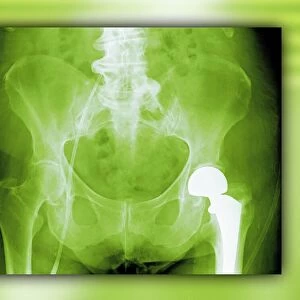
Double hip replacement. Coloured X-ray of the pelvis of a 63 year old male patient who has had two total hip replacements. Each prosthetic hip joint consists of a shaft and ball (red) attached to the top of the femur (thigh bone) and a socket (brown and orange semi-circle) that is inserted into the pelvis. The majority of hip replacements are a result of osteoarthritis, a degenerative joint disease that results in the loss of cartilage between the joint and the growth of bone in place of the cartilage. This causes pain, stiffness and loss of mobility
Science Photo Library features Science and Medical images including photos and illustrations
Media ID 9245721
© ZEPHYR/SCIENCE PHOTO LIBRARY
Artificial Bilateral Double Femur Front Hips Joint Orthopaedics Orthopedics Patient Pelvic Prosthesis Prosthetic Radiography Replaced Sixties Socket Thigh Bone Total Hip Replacement Treatment X Ray Machine Xray Pelvis Sixty Three
FEATURES IN THESE COLLECTIONS
EDITORS COMMENTS
This print showcases a double hip replacement, revealing the intricate details of an artificial joint within the pelvis of a 63-year-old male patient. The vibrant colors highlight the prosthetic components used in this life-changing procedure. Each hip joint consists of a red shaft and ball attached to the top of the femur, while a brown and orange semi-circle socket is inserted into the pelvis. Hip replacements are commonly performed due to osteoarthritis, a degenerative joint disease that causes cartilage loss and bone growth in its place. This painful condition often leads to stiffness and limited mobility. However, through modern medicine and orthopedic expertise, this patient's quality of life has been significantly improved. The image not only captures medical advancements but also represents hope for those suffering from similar conditions. It serves as a reminder that age should never hinder one's ability to seek treatment or regain their independence. With its focus on healthcare, science, and human resilience, this photograph by ZEPHYR/SCIENCE PHOTO LIBRARY encapsulates both artistry and scientific precision. It invites viewers to appreciate how technology can enhance our lives by restoring functionality where it was once lost – ultimately empowering individuals like this resilient 63-year-old man who now stands tall with his new hips.
MADE IN AUSTRALIA
Safe Shipping with 30 Day Money Back Guarantee
FREE PERSONALISATION*
We are proud to offer a range of customisation features including Personalised Captions, Color Filters and Picture Zoom Tools
SECURE PAYMENTS
We happily accept a wide range of payment options so you can pay for the things you need in the way that is most convenient for you
* Options may vary by product and licensing agreement. Zoomed Pictures can be adjusted in the Cart.