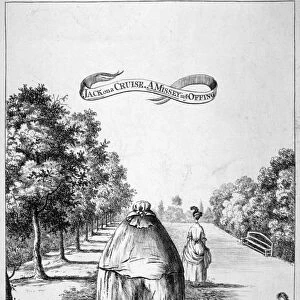
Green fluorescent protein, computer model

![]()
Wall Art and Photo Gifts from Science Photo Library
Green fluorescent protein, computer model
Green fluorescent protein, computer model. This protein is found in the jellyfish Aequorea victoria. When ultraviolet or blue light shines on the protein, it emits green light. This property has made GFP a popular research tool in genetic experiments. The gene that codes for the protein can be introduced to the genome of any animal, plant or fungus, and the cells and tissues that express it will glow green under blue or UV light. Many such " glow in the dark" organisms have been created, including pigs, rabbits and mice. In this model, the secondary structure of the protein is shown. The flat green arrows represent beta sheet regions, the central blue region is an alpha helix
Science Photo Library features Science and Medical images including photos and illustrations
Media ID 6279731
© DR TIM EVANS/SCIENCE PHOTO LIBRARY
Beta Sheet Compound Compounds Green Fluorescent Protein Jelly Fish Marker Molecules Proteins Secondary Structure Sheets Aequorea Victoria Bio Chemistry Biochemical Computer Artwork Genetics Molecular Molecular Model Protein
EDITORS COMMENTS
This print showcases a computer model of the Green Fluorescent Protein (GFP), derived from the mesmerizing jellyfish Aequorea victoria. Known for its remarkable ability to emit green light when exposed to ultraviolet or blue light, GFP has become an invaluable tool in genetic experiments. The gene responsible for this protein can be introduced into the genome of various organisms, including animals, plants, and fungi. Once expressed, these modified cells and tissues will radiate a vibrant green glow under UV or blue light. The significance of GFP extends beyond its natural occurrence as scientists have harnessed its illuminating properties to create numerous "glow in the dark" organisms like pigs, rabbits, and mice. This groundbreaking achievement has revolutionized research in genetics by enabling scientists to track specific genes or proteins within living systems with unparalleled precision. In this stunning computer model representation, we witness the intricate secondary structure of GFP. The flat green arrows elegantly depict beta sheet regions while a central blue region represents an alpha helix. This molecular artwork provides us with a glimpse into the complex world of biochemistry and highlights how scientific advancements continue to unravel nature's mysteries at a molecular level. Captured by Science Photo Library, this image serves as a testament to mankind's relentless pursuit of knowledge and our ability to harness nature's wonders for transformative scientific breakthroughs.
MADE IN AUSTRALIA
Safe Shipping with 30 Day Money Back Guarantee
FREE PERSONALISATION*
We are proud to offer a range of customisation features including Personalised Captions, Color Filters and Picture Zoom Tools
SECURE PAYMENTS
We happily accept a wide range of payment options so you can pay for the things you need in the way that is most convenient for you
* Options may vary by product and licensing agreement. Zoomed Pictures can be adjusted in the Cart.