Insulin molecule C014 / 2290

![]()
Wall Art and Photo Gifts from Science Photo Library
Insulin molecule C014 / 2290
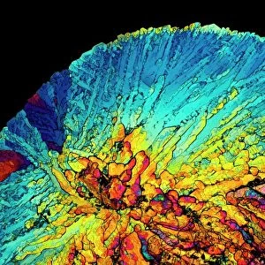
Insulin molecule. Molecular module of insulin showing its primary structure over a background of the molecules electron density map. This map was produced by the British chemist Dorothy Hodgkin (1910-1994) from X-ray crystallography data. Insulin is a hormone produced by the pancreas. It consists of two peptide chains, A and B, which are linked by disulphide bridges (yellow). This is the monomer form of the hormone, the active form, which circulates in the blood. Insulin is released from the pancreas when blood sugar levels are high, for example after a meal. Insufficient production of insulin leads to an accumulation of glucose in the blood causing diabetes
Science Photo Library features Science and Medical images including photos and illustrations
Media ID 9227123
© RAMON ANDRADE 3DCIENCIA/SCIENCE PHOTO LIBRARY
Amino Acid Blood Bridges Compound Compounds Diabetes Diabetes Mellitus Disulphide Bridge Endocrine Endocrinology Hormone Insulin Metabolic Disorder Monomer Pancreatic Primary Structure X Ray Crystallography Biochemical Biochemistry Molecular Molecular Model Protein
EDITORS COMMENTS
This print showcases the intricate structure of the insulin molecule, providing a glimpse into its primary composition. Against a backdrop of an electron density map, meticulously crafted by renowned British chemist Dorothy Hodgkin, this artwork offers a visual representation of the molecular module. Insulin, a vital hormone produced by the pancreas, plays a crucial role in regulating blood sugar levels. Comprised of two peptide chains labeled A and B, these chains are interconnected through vibrant yellow disulphide bridges. This monomer form represents the active state that circulates within our bloodstream. The release of insulin is triggered when blood sugar levels rise after consuming meals. However, insufficient production can lead to diabetes mellitus - a metabolic disorder characterized by elevated glucose accumulation in the blood. Through X-ray crystallography data and Hodgkin's expertise, we gain insight into this complex biochemical compound responsible for maintaining our body's equilibrium. The detailed illustration highlights amino acids and protein structures essential for understanding insulin's function within the endocrine system. Ramon Andrade from 3DCIENCIA/SCIENCE PHOTO LIBRARY has skillfully captured both artistry and scientific precision in this remarkable piece. It serves as a reminder of how advancements in biochemistry enable us to comprehend fundamental processes occurring within our bodies while appreciating their inherent beauty.
MADE IN AUSTRALIA
Safe Shipping with 30 Day Money Back Guarantee
FREE PERSONALISATION*
We are proud to offer a range of customisation features including Personalised Captions, Color Filters and Picture Zoom Tools
SECURE PAYMENTS
We happily accept a wide range of payment options so you can pay for the things you need in the way that is most convenient for you
* Options may vary by product and licensing agreement. Zoomed Pictures can be adjusted in the Cart.