Jigsaw Puzzle > Science > Xray
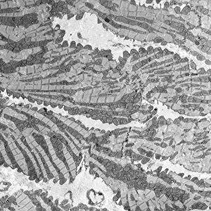
Jigsaw Puzzle : Healthy knee, CT scan C018 / 0413


![]()
Jigsaw Puzzles from Science Photo Library
Healthy knee, CT scan C018 / 0413
Healthy knee. Coloured frontal computed tomography (CT) scan projected on to a magnetic resonance imaging (MRI) scan of the knee of a 30 year old. The knee joint is formed by the articulation of the femur (thigh bone, top) with tibia (shin bone, bottom centre). The smaller fibula (bottom left) is also seen. The patella (knee cap) is seen over the bottom of the femur
Science Photo Library features Science and Medical images including photos and illustrations
Media ID 9237677
© ZEPHYR/SCIENCE PHOTO LIBRARY
Calf Bone Composite Ct Scan Femur Fibula Front Frontal Joint Knee Knee Cap Lower Limb Mri Scan Patella Radiography Scanner Shin Bone Thigh Bone Thirties Thirty Tibia X Ray Machine Xray
Jigsaw Puzzle (500 Pieces)
Discover the fascinating intersection of health and technology with Media Storehouse's Jigsaw Puzzle collection. This captivating puzzle, featuring a unique combination of a healthy knee and a CT scan image from ZEPHYR/SCIENCE PHOTO LIBRARY, provides an intriguing look at the advanced medical imaging technology used to examine our bodies. Piece together the intricate details of this colorful and educational puzzle, perfect for puzzle enthusiasts, healthcare professionals, or anyone with an interest in science and human anatomy.
500 piece puzzles are custom made in Australia and hand-finished on 100% recycled 1.6mm thick laminated puzzle boards. There is a level of repetition in jigsaw shapes with each matching piece away from its pair. The completed puzzle measures 40x51cm and is delivered packaged in an attractive presentation box specially designed to fit most mail slots with a unique magnetic lid
Jigsaw Puzzles are an ideal gift for any occasion
Estimated Product Size is 50.7cm x 40.3cm (20" x 15.9")
These are individually made so all sizes are approximate
Artwork printed orientated as per the preview above, with landscape (horizontal) or portrait (vertical) orientation to match the source image.
FEATURES IN THESE COLLECTIONS
EDITORS COMMENTS
This print showcases a healthy knee in all its intricate glory. The image, titled "Healthy knee, CT scan C018 / 0413" offers a coloured frontal computed tomography (CT) scan projected onto a magnetic resonance imaging (MRI) scan of the knee of a vibrant 30-year-old individual. The joint responsible for supporting this remarkable limb is formed by the articulation of the femur, or thigh bone, positioned at the top, with the tibia, or shin bone, located at the bottom center. Additionally, we catch sight of the smaller fibula on the bottom left side. Overlying the base of the femur is our familiar friend -the patella- commonly known as our knee cap. This composite radiographic masterpiece provides an extraordinary glimpse into human anatomy and biology. It serves as a reminder that within each person lies an incredible network of bones and joints working harmoniously to facilitate movement and mobility. Expertly captured through cutting-edge technology such as MRI scans and CT scans using X-ray machines, this image highlights both beauty and functionality simultaneously. With its vivid colors and precise details revealing every contour and structure within this lower limb marvelously. ZEPHYR/SCIENCE PHOTO LIBRARY has once again delivered an awe-inspiring visual representation that not only educates but also captivates viewers with its sheer brilliance in showcasing normal anatomical features found within our bodies.
MADE IN AUSTRALIA
Safe Shipping with 30 Day Money Back Guarantee
FREE PERSONALISATION*
We are proud to offer a range of customisation features including Personalised Captions, Color Filters and Picture Zoom Tools
SECURE PAYMENTS
We happily accept a wide range of payment options so you can pay for the things you need in the way that is most convenient for you
* Options may vary by product and licensing agreement. Zoomed Pictures can be adjusted in the Cart.