Trichophyton fungus, SEM

![]()
Wall Art and Photo Gifts from Science Photo Library
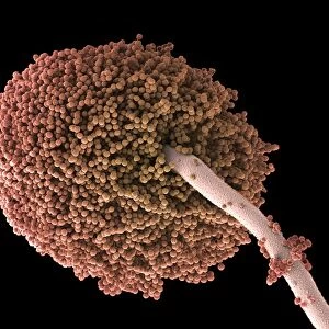
Trichophyton fungus, SEM
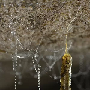
Trichophyton fungus. Coloured scanning electron micrograph (SEM) of a dermatophyte fungus belonging to the genus Trichophyton. The thread-like structures are hyphae, the vegetative part of the fungus. Dermatophyte fungi infect keratinised tissues (skin, nails, hair) in humans and other animals
Science Photo Library features Science and Medical images including photos and illustrations
Media ID 6275984
© SUSUMU NISHINAGA/SCIENCE PHOTO LIBRARY
Brown Conditions Dermatophyte Diseases False Colour Filamentous Fungal Fungi Fungus Hypha Hyphae Infection Infectious Micro Organism Microbe Mycelium Mycology Pathogenic Condition Dermatophytosis Disorder False Coloured Micro Biology Microbiological Mono Chrome Pathogen Tinea
EDITORS COMMENTS
This print showcases the intricate beauty of a Trichophyton fungus, captured through a scanning electron microscope (SEM). The false-colored image highlights the various structures and features of this dermatophyte fungus, which belongs to the genus Trichophyton. The thread-like formations visible in the photograph are known as hyphae, representing the vegetative part of the fungus. Trichophyton fungi primarily infect keratinized tissues such as skin, nails, and hair in both humans and other animals. This makes them responsible for causing conditions like tinea or dermatophytosis. Through its rich brown hues and detailed composition, this SEM image offers a glimpse into the world of microbiology. It reveals how pathogenic microorganisms can manifest themselves on a cellular level, emphasizing their potential to cause infectious disorders. As we delve into mycology and explore fungal biology further, images like these serve as invaluable tools for research purposes. They help scientists study pathogens at an incredibly detailed level while providing insights into diseases caused by dermatophytes. This particular print is courtesy of Science Photo Library—a trusted source for scientific imagery—showcasing their commitment to capturing visually stunning representations of nature's wonders. While it may not be intended for commercial use specifically mentioned by Science Photo Library guidelines, it undoubtedly serves as an educational resource that sparks curiosity about our complex biological world.
MADE IN AUSTRALIA
Safe Shipping with 30 Day Money Back Guarantee
FREE PERSONALISATION*
We are proud to offer a range of customisation features including Personalised Captions, Color Filters and Picture Zoom Tools
SECURE PAYMENTS
We happily accept a wide range of payment options so you can pay for the things you need in the way that is most convenient for you
* Options may vary by product and licensing agreement. Zoomed Pictures can be adjusted in the Cart.