
Canvas Print : Hip bones, artwork C016 / 7015


![]()
Canvas Prints from Science Photo Library
Hip bones, artwork C016 / 7015
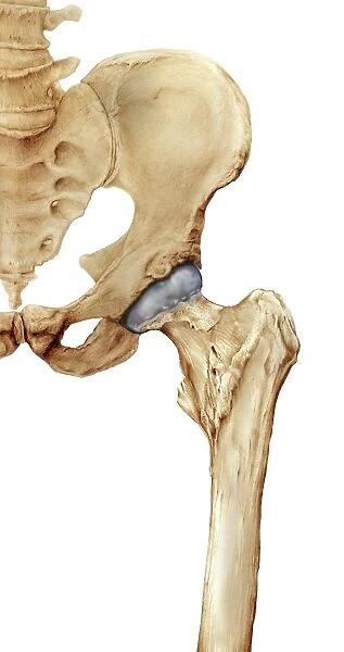
Hip bones. Artwork of a frontal (anterior) view of the left hip, showing the bones that articulate to form this joint. At lower right is the femur (thigh bone). The head of the femur (ball-shaped) is covered in cartilage (grey) that cushions it as it moves in the acetabulum, the hip socket in the pelvis. The pelvis bones that form part of the hip joint are the ischium and pubis (centre left) and the ilium (upper centre). At top left are the lower bones of the spine, the tailbone (coccyx), sacrum, and several lumbar vertebrae
Science Photo Library features Science and Medical images including photos and illustrations
Media ID 9245961
© D & L GRAPHICS / SCIENCE PHOTO LIBRARY
Acetabulum Anterior Articulation Backbone Bones Cartilage Coccyx Femoral Femoral Head Femur Frontal Hip Socket Ilium Ischium Joint Lumbar Pelvic Pubis Sacrum Tail Bone Tailbone Thigh Bone Pelvis Vertebrae
30"x20" (76x51cm) Canvas Print
Add an element of anatomy art to your home decor with our Canvas Prints from Media Storehouse. This captivating artwork, titled "Hip bones, artwork C016 / 7015" by D & L Graphics / Science Photo Library, showcases a detailed and intriguing frontal view of the left hip, highlighting the articulating bones including the femur (thigh bone). Ideal for those with an appreciation for science and anatomy, this stunning print brings a unique touch to any room. Order now and bring the beauty of the human body into your living space.
Delivered stretched and ready to hang our premium quality canvas prints are made from a polyester/cotton blend canvas and stretched over a 1.25" (32mm) kiln dried knot free wood stretcher bar. Packaged in a plastic bag and secured to a cardboard insert for safe transit.
Canvas Prints add colour, depth and texture to any space. Professionally Stretched Canvas over a hidden Wooden Box Frame and Ready to Hang
Estimated Image Size (if not cropped) is 40.2cm x 76.2cm (15.8" x 30")
Estimated Product Size is 50.8cm x 76.2cm (20" x 30")
These are individually made so all sizes are approximate
Artwork printed orientated as per the preview above, with portrait (vertical) orientation to match the source image.
EDITORS COMMENTS
This print titled "Hip bones, artwork C016 / 7015" offers a detailed frontal view of the left hip, showcasing the intricate bones that come together to form this vital joint. The image highlights the femur, or thigh bone, positioned at the lower right corner. Its ball-shaped head is covered in grey cartilage, providing cushioning as it moves within the acetabulum - the hip socket located in the pelvis. The pelvic bones involved in this joint are prominently displayed at center-left and upper-center. These include the ischium and pubis, forming part of the hip joint along with the ilium situated above them. At top left, we can observe several lumbar vertebrae alongside other lower spine components such as sacrum and coccyx (tailbone). This illustration on a white background not only showcases anatomical accuracy but also emphasizes elements related to biology and human anatomy. It provides valuable insight into how our hips function normally while highlighting key structures like vertebrae and leg bones. Created by D & L GRAPHICS for Science Photo Library, this artwork serves as an educational resource for those interested in understanding hip joints from a scientific perspective.
MADE IN AUSTRALIA
Safe Shipping with 30 Day Money Back Guarantee
FREE PERSONALISATION*
We are proud to offer a range of customisation features including Personalised Captions, Color Filters and Picture Zoom Tools
SECURE PAYMENTS
We happily accept a wide range of payment options so you can pay for the things you need in the way that is most convenient for you
* Options may vary by product and licensing agreement. Zoomed Pictures can be adjusted in the Cart.