Photo Mug > Arts > Realistic drawings > Nature art > Botanical artwork
Photo Mug : Diatom alga, SEM


![]()
Home Decor from Science Photo Library
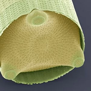
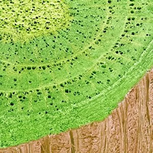
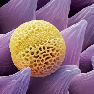
Diatom alga, SEM
Diatom. Coloured scanning electron micrograph (SEM) of a Biddulphia sp. diatom. This is a marine planktonic unicellular alga. It has a mineralised cell wall (frustule) divided into two halves. The frustule contains silica and provides protection and support
Science Photo Library features Science and Medical images including photos and illustrations
Media ID 6276653
© STEVE GSCHMEISSNER/SCIENCE PHOTO LIBRARY
Alga Algae Algal Diatom Frustule Phytoplankton Plankton Planktonic Single Celled Unicellular False Coloured
Photo Mug
Bring the wonders of the marine world into your daily routine with Media Storehouse's Photo Mugs. Featuring an exquisite Coloured Scanning Electron Micrograph (SEM) image of a Biddulphia sp. diatom from Science Photo Library, this mug showcases the intricate details of this marine planktonic unicellular alga's mineralised cell wall (frustule), divided into two halves. Each sip from this unique mug is a reminder of the beauty and complexity of nature. Perfect for scientists, marine enthusiasts, or anyone who appreciates the wonders of the natural world. Embrace the power of a picture to brighten your day, every day!
A personalised photo mug blends sentimentality with functionality, making an ideal gift for cherished loved ones, close friends, or valued colleagues. Preview may show both sides of the same mug.
Elevate your coffee or tea experience with our premium white ceramic mug. Its wide, comfortable handle makes drinking easy, and you can rely on it to be both microwave and dishwasher safe. Sold in single units, preview may show both sides of the same mug so you can see how the picture wraps around.
Mug Size is 8.1cm high x 9.6cm diameter (3.2" x 3.8")
These are individually made so all sizes are approximate
FEATURES IN THESE COLLECTIONS
> Arts
> Realistic drawings
> Nature art
> Botanical artwork
EDITORS COMMENTS
This print showcases the intricate beauty of a Diatom alga, captured through a coloured scanning electron microscope (SEM). The image reveals a Biddulphia sp. diatom, an enchanting marine planktonic unicellular alga that thrives in our oceans. What makes this diatom truly remarkable is its mineralized cell wall, known as a frustule, which is divided into two halves. Composed primarily of silica, this frustule not only offers protection to the delicate organism but also provides essential support for its survival. The vibrant colours portrayed in this photograph are false-coloured enhancements added during the imaging process to highlight specific features and characteristics of the diatom. Through advanced technology and scientific expertise, we can now appreciate the mesmerizing details that would otherwise remain hidden from our naked eyes. As we delve into nature's botanical wonders, it becomes evident how diverse and awe-inspiring life on Earth truly is. This single-celled marvel serves as a reminder of the extraordinary complexity found within even seemingly simple organisms. With each glance at this photo print, one cannot help but be captivated by the elegance and intricacy displayed by this marine phytoplankton. It stands as a testament to both the power of biology and mankind's ability to unlock nature's secrets through cutting-edge tools like scanning electron microscopes.
MADE IN AUSTRALIA
Safe Shipping with 30 Day Money Back Guarantee
FREE PERSONALISATION*
We are proud to offer a range of customisation features including Personalised Captions, Color Filters and Picture Zoom Tools
SECURE PAYMENTS
We happily accept a wide range of payment options so you can pay for the things you need in the way that is most convenient for you
* Options may vary by product and licensing agreement. Zoomed Pictures can be adjusted in the Cart.