Photo Mug : Purkinje nerve cell


![]()
Home Decor from Science Photo Library
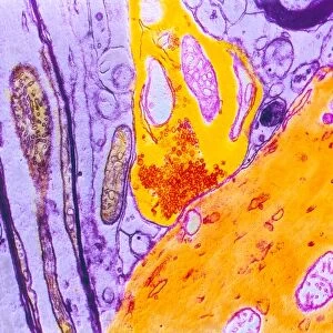
Purkinje nerve cell
Science Photo Library features Science and Medical images including photos and illustrations
Media ID 6421844
© DAVID BECKER/SCIENCE PHOTO LIBRARY
Cerebellum Confocal Dendrite Dendrites Fluorescence Fluorescent Granular Grey Matter Histology Immunofluorescence Immunofluorescent Layers Molecular Layer Nerve Cell Nervous Neuron Neurone Purkinje System Tissue Brain Cells Light Micrograph Neurology
Photo Mug
Bring the beauty of science into your daily routine with Media Storehouse Photo Mugs. Featuring an exquisite image of a Purkinje nerve cell from Science Photo Library, these mugs are not just functional, but also a conversation starter. Each mug showcases high-quality, vibrant prints that capture the intricacy and detail of the scientific world. Perfect for scientists, students, or anyone with a passion for biology, these mugs make a unique and thoughtful gift. Sip your favorite beverage while marveling at the complexity of the natural world, one cell at a time.
A personalised photo mug blends sentimentality with functionality, making an ideal gift for cherished loved ones, close friends, or valued colleagues. Preview may show both sides of the same mug.
Elevate your coffee or tea experience with our premium white ceramic mug. Its wide, comfortable handle makes drinking easy, and you can rely on it to be both microwave and dishwasher safe. Sold in single units, preview may show both sides of the same mug so you can see how the picture wraps around.
Mug Size is 9.6cm high x 8.1cm diameter (3.8" x 3.2")
These are individually made so all sizes are approximate
EDITORS COMMENTS
This print showcases the intricate beauty of a Purkinje nerve cell, also known as a Purkinje neuron. The image provides an up-close look at this vital component of our nervous system, revealing its complex structure and function. The Purkinje nerve cell is found in the cerebellum, which plays a crucial role in coordinating movement and balance. Its distinctive shape and branching dendrites are highlighted by fluorescent labeling techniques used in histology research. This immunofluorescent staining allows scientists to visualize specific molecules within the cell, providing valuable insights into its functioning. The vibrant fluorescence brings to life the molecular layer surrounding the grey matter of the cerebellum. Each individual layer represents different populations of cells that work together to transmit electrical signals throughout the brain. This stunning light micrograph not only serves as a visual feast for biology enthusiasts but also holds great significance for neurology researchers studying various aspects of brain function and health. By understanding how these neurons communicate with each other, scientists can gain deeper insights into neurological disorders such as Parkinson's disease or stroke recovery. Science Photo Library has once again captured an awe-inspiring moment from nature's own laboratory, reminding us of both the complexity and elegance present within our own bodies.
MADE IN AUSTRALIA
Safe Shipping with 30 Day Money Back Guarantee
FREE PERSONALISATION*
We are proud to offer a range of customisation features including Personalised Captions, Color Filters and Picture Zoom Tools
SECURE PAYMENTS
We happily accept a wide range of payment options so you can pay for the things you need in the way that is most convenient for you
* Options may vary by product and licensing agreement. Zoomed Pictures can be adjusted in the Cart.