Cytoskeleton, TEM

![]()
Wall Art and Photo Gifts from Science Photo Library
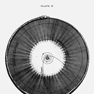
Cytoskeleton, TEM
Cytoskeleton. Coloured transmission electron micrograph (TEM) of the cytoskeleton of a human skin cell. The cell nucleus is at centre right. The rest of the cells contents have been biochemically extracted. The cytoskeleton is made up of three types of proteins; actin filaments, microtubules and intermediate filaments. The cytoskeleton maintains the cells shape, allows some cellular mobility and is involved in intracellular transport. Magnification: x3, 800 when printed at 10 centimetres wide
Science Photo Library features Science and Medical images including photos and illustrations
Media ID 6321681
© SCIENCE PHOTO LIBRARY
Actin Biochemical Extraction Cell Biology Cell Shape Colored Cytological Cytology Cytoskeletal Cytoskeleton Filament Human Skin Cell Microfilaments Microscope Microtubule Nucleus Organelle Transmission Electron Transmission Electron Micrograph Tubules Tubulin Blue Background Protein
EDITORS COMMENTS
This print from Science Photo Library showcases the intricate beauty of a human skin cell's cytoskeleton. In this coloured transmission electron micrograph (TEM), we are granted a glimpse into the complex inner workings of cellular biology. At the centre right, we can see the cell nucleus, surrounded by an array of vibrant structures. These structures make up the cytoskeleton, which consists of three types of proteins: actin filaments, microtubules, and intermediate filaments. The cytoskeleton plays multiple crucial roles within the cell; it maintains its shape, enables cellular mobility, and facilitates intracellular transport. The blue background provides a striking contrast to highlight these microscopic wonders. Through biochemical extraction techniques employed in capturing this image, all other contents of the cell have been removed except for its structural framework. Printed at 10 centimetres wide with a magnification level of x3,800, every detail is meticulously preserved for our exploration and admiration. This photograph not only serves as an artistic masterpiece but also offers valuable insights into one aspect of cellular life that often goes unnoticed. In summary, Science Photo Library has once again provided us with an awe-inspiring visual representation that bridges science and art seamlessly.
MADE IN AUSTRALIA
Safe Shipping with 30 Day Money Back Guarantee
FREE PERSONALISATION*
We are proud to offer a range of customisation features including Personalised Captions, Color Filters and Picture Zoom Tools
SECURE PAYMENTS
We happily accept a wide range of payment options so you can pay for the things you need in the way that is most convenient for you
* Options may vary by product and licensing agreement. Zoomed Pictures can be adjusted in the Cart.