Home > Science > SEM
Diatom alga, SEM

![]()
Wall Art and Photo Gifts from Science Photo Library
Diatom alga, SEM
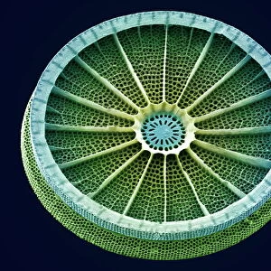
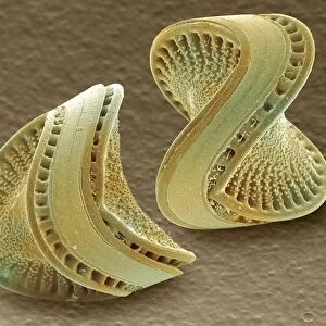
Diatom. Coloured scanning electron micrograph (SEM) of a Skeletonema punctatum diatom. This is a planktonic unicellular alga. It has a mineralised cell wall (frustule) divided into two halves. The frustule contains silica and provides protection and support. Magnification: x85 at 6x7cm size
Science Photo Library features Science and Medical images including photos and illustrations
Media ID 6292483
© STEVE GSCHMEISSNER/SCIENCE PHOTO LIBRARY
Alga Algae Algal Centric Cylinder Cylindrical Diatom Diatoms Frustule Magnified Image Microscopic Photos Phytoplankton Plankton Planktonic Radial Symmetry Single Celled Subjects Symmetrical Unicellular
MADE IN AUSTRALIA
Safe Shipping with 30 Day Money Back Guarantee
FREE PERSONALISATION*
We are proud to offer a range of customisation features including Personalised Captions, Color Filters and Picture Zoom Tools
SECURE PAYMENTS
We happily accept a wide range of payment options so you can pay for the things you need in the way that is most convenient for you
* Options may vary by product and licensing agreement. Zoomed Pictures can be adjusted in the Cart.