Canvas Print : Rod and cone cells of the eye, SEM C014 / 4865


![]()
Canvas Prints from Science Photo Library
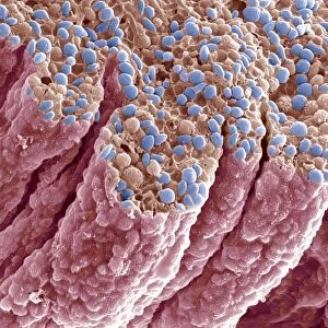
Rod and cone cells of the eye, SEM C014 / 4865
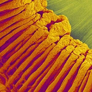
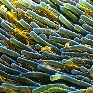
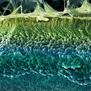
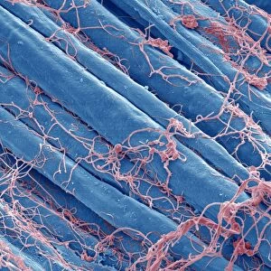
Rod and cone cells of the eye. Coloured scanning electron micrograph (SEM) of rod and cone cells in the retina of a mammalian eye. Cone cells and the more numerous rod cells are specialised light-sensitive cells that occur on the surface of the retina. They are responsible for detecting visible images, which are transmitted as nerve impulses to the optic nerve and the brain. There are about 130 million rod cells in the human retina, which detect light intensity and so are important for day and night vision. The less numerous cone-like cone cells (about 6.5 million in the human retina) respond specifically to colour. Magnification: x5, 300 when printed at 10 centimetres wide
Science Photo Library features Science and Medical images including photos and illustrations
Media ID 9225701
© CLOUDS HILL IMAGING LTD/SCIENCE PHOTO LIBRARY
Colored Cone Cell Cones Light Sensitive Mammal Mammalian Neural Neuron Neuronal Photoreceptor Photoreceptors Receptor Receptors Reptile Retina Retinal Rods Sense Sensory Sight Vision Visual Cells Nervous System
20"x16" (51x41cm) Canvas Print
Bring the wonders of science into your home with Media Storehouse's Canvas Prints. This captivating image, "Rod and Cone Cells of the Eye" (CLOUDS HILL IMAGING LTD/SCIENCE PHOTO LIBRARY/C014 / 4865), showcases the intricate beauty of the human eye in vivid detail. Colored Scanning Electron Micrograph (SEM) reveals the complex structure of rod and cone cells in the retina, essential for vision in different lighting conditions. Elevate your space with this striking, high-quality canvas print and ignite curiosity in the world around us.
Delivered stretched and ready to hang our premium quality canvas prints are made from a polyester/cotton blend canvas and stretched over a 1.25" (32mm) kiln dried knot free wood stretcher bar. Packaged in a plastic bag and secured to a cardboard insert for safe transit.
Canvas Prints add colour, depth and texture to any space. Professionally Stretched Canvas over a hidden Wooden Box Frame and Ready to Hang
Estimated Product Size is 50.8cm x 40.6cm (20" x 16")
These are individually made so all sizes are approximate
Artwork printed orientated as per the preview above, with landscape (horizontal) orientation to match the source image.
EDITORS COMMENTS
This print showcases the intricate beauty of rod and cone cells in the eye. In this coloured scanning electron micrograph (SEM), we are granted a glimpse into the remarkable world of these specialized light-sensitive cells that reside on the surface of the retina. Rod and cone cells play a vital role in our visual perception, as they are responsible for detecting visible images and transmitting them as nerve impulses to the optic nerve and ultimately to our brain. With approximately 130 million rod cells, which detect light intensity, and around 6.5 million cone-like cells that respond specifically to colour, our eyes possess an astonishing capacity for day and night vision. The magnified view reveals their delicate structures with astounding clarity. Each cell is meticulously designed to fulfill its unique function within our visual system. The rods outnumbering cones significantly highlight their importance in low-light conditions, while cones contribute to our ability to perceive vibrant colors. This image not only highlights the complexity of these sensory receptors but also emphasizes their crucial role in enabling us to experience the wonders of nature's palette firsthand. It serves as a reminder of how intricately woven biology is with every aspect of life – from wildlife exploration to understanding human anatomy. Captured by CLOUDS HILL IMAGING LTD/SCIENCE PHOTO LIBRARY using a scanning electron microscope (SEM), this photograph stands as a testament to both scientific advancement and artistic appreciation for nature's marvels.
MADE IN AUSTRALIA
Safe Shipping with 30 Day Money Back Guarantee
FREE PERSONALISATION*
We are proud to offer a range of customisation features including Personalised Captions, Color Filters and Picture Zoom Tools
SECURE PAYMENTS
We happily accept a wide range of payment options so you can pay for the things you need in the way that is most convenient for you
* Options may vary by product and licensing agreement. Zoomed Pictures can be adjusted in the Cart.