Cushion : Left hip and nerve plexus, artwork C016 / 6808


![]()
Home Decor from Science Photo Library
Left hip and nerve plexus, artwork C016 / 6808
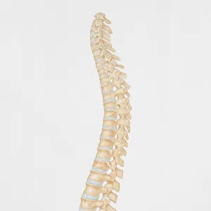
Left hip and nerve plexus. Artwork of the nerves (yellow) and bones of the left hip, seen from the front. This is the lumbar plexus, itself forming part of the lumbosacral plexus, including lumbar and sacral nerves. The main hip nerves present here are those shown passing over or close to the head of the femur. From left to right, these are: the sciatic nerve, the femoral nerve, and the lateral femoral cutaneous nerve. For the right hip, see C016/6809
Science Photo Library features Science and Medical images including photos and illustrations
Media ID 9246219
© D & L GRAPHICS / SCIENCE PHOTO LIBRARY
Anterior Backbone Bones Bundle Femoral Femoral Nerve Femur Frontal Iliac Joint Lower Back Lumbar Plexus Lumbosacral Plexus Nerve Nerve Plexus Nerves Neural Pelvic Sacral Sacrum Sciatic Nerve Skeletal Vertebral Column Neurological Neurology Pelvis
Cushion
Refresh your home decor with a beautiful full photo 16"x16" (40x40cm) cushion, complete with cushion pad insert. Printed on both sides and made from 100% polyester with a zipper on the bottom back edge of the cushion cover. Care Instructions: Warm machine wash, do not bleach, do not tumble dry. Warm iron inside out. Do not dry clean.
Accessorise your space with decorative, soft cushions
Estimated Product Size is 40cm x 40cm (15.7" x 15.7")
These are individually made so all sizes are approximate
Artwork printed orientated as per the preview above, with landscape (horizontal) or portrait (vertical) orientation to match the source image.
EDITORS COMMENTS
This print showcases the intricate network of nerves and bones in the left hip region. The artwork, titled "Left hip and nerve plexus" provides a detailed illustration of the lumbar plexus, which is part of the lumbosacral plexus encompassing both lumbar and sacral nerves. From a frontal perspective, we can observe the yellow-colored nerves intertwining with the skeletal structure. The main focus lies on three prominent hip nerves that either pass over or closely associate with the femur's head. These significant nerves are identified from left to right as follows: sciatic nerve, femoral nerve, and lateral femoral cutaneous nerve. This comprehensive depiction not only highlights their anatomical positions but also emphasizes their vital role in transmitting neural signals throughout this region. The white background enhances our focus on this remarkable artwork while emphasizing its scientific nature. By exploring this image, one gains insight into various aspects such as joint health, spine anatomy, normal skeletal structure, and neurological connections within the human body. Created by D & L GRAPHICS for Science Photo Library (not associated with any company), this illustrative masterpiece serves as an invaluable resource for professionals in biology-related fields like neurology or orthopedics. It offers a deeper understanding of bone structures like pelvis and vertebral column along with key elements like sacrum and lower back. In summary, this visually striking print captures both beauty and complexity through its portrayal of left hip anatomy intertwined with neural pathways—a true testament
MADE IN AUSTRALIA
Safe Shipping with 30 Day Money Back Guarantee
FREE PERSONALISATION*
We are proud to offer a range of customisation features including Personalised Captions, Color Filters and Picture Zoom Tools
SECURE PAYMENTS
We happily accept a wide range of payment options so you can pay for the things you need in the way that is most convenient for you
* Options may vary by product and licensing agreement. Zoomed Pictures can be adjusted in the Cart.