Cushion : Multi-celled embryo, artwork
![]()

Home Decor from Science Photo Library
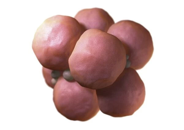
Multi-celled embryo, artwork
Multi-celled embryo. Image 4 of 4. Computer model representing a cluster of daughter cells. The development of an embryo is called embryogenesis. In organisms that reproduce sexually, once a sperm fertilizes an egg cell, the result is a cell that has all the DNA of the two parents (zygote). The zygote begins to divide by mitosis producing 2, 4, 6, 8 cells and so on, to form a multicellular organism. The result of this process is an embryo. For a sequence representing mitosis and embryo development see images P680/0943-P680/0946
Science Photo Library features Science and Medical images including photos and illustrations
Media ID 6454873
© DAVID MACK/SCIENCE PHOTO LIBRARY
Beginning Cell Biology Cytology Daughter Cell Daughter Cells Development Developmental Biology Embryo Embryology Life Mitosis Mitotic Origin Physiological Physiology Re Production Series Start Cells Embryogenesis
Cushion
Refresh your home decor with a beautiful full photo 16"x16" (40x40cm) cushion, complete with cushion pad insert. Printed on both sides and made from 100% polyester with a zipper on the bottom back edge of the cushion cover. Care Instructions: Warm machine wash, do not bleach, do not tumble dry. Warm iron inside out. Do not dry clean.
Accessorise your space with decorative, soft cushions
Estimated Product Size is 40cm x 40cm (15.7" x 15.7")
These are individually made so all sizes are approximate
Artwork printed orientated as per the preview above, with landscape (horizontal) or portrait (vertical) orientation to match the source image.
EDITORS COMMENTS
This print showcases the intricate beauty of a multi-celled embryo, depicted through an exquisite computer model. Embryogenesis, the process of embryo development, is a remarkable phenomenon in sexually reproducing organisms. It commences when a sperm successfully fertilizes an egg cell, resulting in a zygote that carries the combined DNA of both parents. The image portrays a cluster of daughter cells emerging from this initial zygote through mitosis – the process by which cells divide and multiply. As each division occurs, the number of cells increases exponentially; 2 becomes 4, then 6, then 8 and so forth until they form a complex multicellular organism. This artwork serves as an illustration for the origins and beginnings of life itself. Its detailed representation highlights various aspects within biology such as reproduction, anatomy, physiology, and developmental biology. The cut-out style adds depth to our understanding of embryology and cytology while emphasizing how multiple physiological processes work harmoniously to shape human bodies. Science Photo Library has masterfully captured this pivotal stage in biological development with their expertise in scientific imagery. This photograph not only educates but also captivates viewers with its artistic portrayal of cellular life's incredible journey from conception to embryo formation.
MADE IN AUSTRALIA
Safe Shipping with 30 Day Money Back Guarantee
FREE PERSONALISATION*
We are proud to offer a range of customisation features including Personalised Captions, Color Filters and Picture Zoom Tools
SECURE PAYMENTS
We happily accept a wide range of payment options so you can pay for the things you need in the way that is most convenient for you
* Options may vary by product and licensing agreement. Zoomed Pictures can be adjusted in the Cart.


