Greetings Card : Uterine fibroid, light micrograph C015 / 6412


![]()
Cards from Science Photo Library
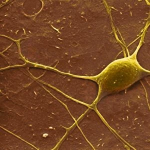
Uterine fibroid, light micrograph C015 / 6412
Uterine fibroid. Light micrograph of a section through a uterine fibroid (oval). A fibroid is a fibrous benign tumour originating from muscular tissue. Most do not require treatment, but some can cause bleeding, pain and other disorders. Large fibroids may need to be surgically removed
Science Photo Library features Science and Medical images including photos and illustrations
Media ID 9208227
© CNRI/SCIENCE PHOTO LIBRARY
Benign Female Reproductive System Fibroid Fibroma Fibrous Growth Gynaecology Gynecology Histopathological Histopathology Leiomyoma Myoma Stain Stained Tumour Uterine Uterus Womb Abnormal Condition Disorder Light Micrograph Light Microscope Section Sectioned Unhealthy
Greetings Card (7"x5")
"Discover the wonders of the human body with our unique range of scientific greetings cards from Media Storehouse. This card features a captivating light micrograph image of a uterine fibroid, showcasing the intricate details of this benign tumor's fibrous structure. A fascinating and educational gift for those with an appreciation for biology and medical science." - Media Storehouse. (Image credit: CNRI/SCIENCE PHOTO LIBRARY - Ref: C015 / 6412)
Folded Greeting Cards (12.5x17.5 cm) have a laminate finish and are supplied with an envelope. The front and inside can be personalised with text in a selection of fonts, layouts and colours.
Greetings Cards suitable for Birthdays, Weddings, Anniversaries, Graduations, Thank You and much more
Estimated Product Size is 12.5cm x 17.5cm (4.9" x 6.9")
These are individually made so all sizes are approximate
Artwork printed orientated as per the preview above, with landscape (horizontal) or portrait (vertical) orientation to match the source image.
EDITORS COMMENTS
This print showcases a light micrograph of a uterine fibroid, providing an intriguing glimpse into the world of medical histopathology. The image captures a section through an oval-shaped fibrous benign tumor originating from muscular tissue in the uterus. Uterine fibroids are commonly found in women and while most do not necessitate treatment, some can lead to complications such as bleeding, pain, and other disorders. The stained micrograph vividly highlights the abnormal growth within the female reproductive system. With its intricate details visible under the light microscope, this image serves as a valuable resource for gynecologists and researchers studying conditions related to the uterus. The significance of this photograph lies in its potential impact on medical advancements and patient care. By understanding the characteristics and behavior of uterine fibroids at a microscopic level, healthcare professionals can make informed decisions regarding treatment options. In cases where large fibroids pose significant health risks or impair quality of life, surgical removal may be necessary. Provided by CNRI/SCIENCE PHOTO LIBRARY, this visually striking print offers both scientific insight and aesthetic appeal. It reminds us of the complexities that exist within our bodies and underscores the importance of ongoing research in gynecology to improve women's health worldwide.
MADE IN AUSTRALIA
Safe Shipping with 30 Day Money Back Guarantee
FREE PERSONALISATION*
We are proud to offer a range of customisation features including Personalised Captions, Color Filters and Picture Zoom Tools
SECURE PAYMENTS
We happily accept a wide range of payment options so you can pay for the things you need in the way that is most convenient for you
* Options may vary by product and licensing agreement. Zoomed Pictures can be adjusted in the Cart.