Cushion > Science > SEM
Cushion : Retina, SEM


![]()
Home Decor from Science Photo Library
Retina, SEM
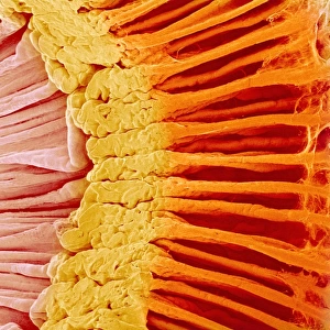
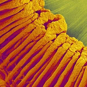
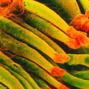
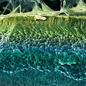
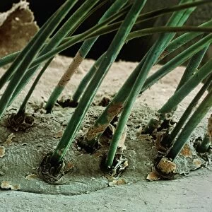
Retina. Coloured scanning electron micrograph (SEM) of a section through a freeze-fractured retina, showing the light-sensitive rods and cones and their associated neurones. Light entering the eyes passes through the bipolar neurones in the nuclear layers (purple, lower frame). It then hits the rods and cones (pink/yellow, upper frame). The rods, used in dim light, are found at the retinal margins. The cones, which control colour vision and acuity (sharpness), function best in bright light. They are concentrated in the centre of the retina. Information from both is passed to the neurons, which relay it to the optic nerve (not seen). Magnification: x1000 at 6x7cm size
Science Photo Library features Science and Medical images including photos and illustrations
Media ID 6449335
© STEVE GSCHMEISSNER/SCIENCE PHOTO LIBRARY
Bipolar Cone Cones Fibre Fibres Fracture Fractured Freeze Fractured Ganglion Histology Layer Layered Layers Light Sensitive Nerve Nervous Neuron Neurone Neurones Neurons Optic Optical Photoreceptor Photoreceptors Receptor Receptors Retina Retinal Rods Sight Tissue Vision Visual Sense Cells Section Sectioned
Cushion
Refresh your home decor with a beautiful full photo 16"x16" (40x40cm) cushion, complete with cushion pad insert. Printed on both sides and made from 100% polyester with a zipper on the bottom back edge of the cushion cover. Care Instructions: Warm machine wash, do not bleach, do not tumble dry. Warm iron inside out. Do not dry clean.
Accessorise your space with decorative, soft cushions
Estimated Product Size is 40cm x 40cm (15.7" x 15.7")
These are individually made so all sizes are approximate
Artwork printed orientated as per the preview above, with landscape (horizontal) or portrait (vertical) orientation to match the source image.
EDITORS COMMENTS
This print from Science Photo Library showcases the intricate beauty of the retina, a vital component of our visual system. In this coloured scanning electron micrograph (SEM), we are granted a glimpse into the complex layers and cells that make up this remarkable tissue. The image reveals an exquisite section through a freeze-fractured retina, highlighting the presence of light-sensitive rods and cones along with their associated neurones. The lower frame illustrates the bipolar neurones in purple, which play a crucial role in transmitting light entering the eyes. Moving to the upper frame, we observe the rods and cones depicted in pink and yellow respectively. These photoreceptor cells have distinct functions - while rods aid us in dim lighting conditions, cones enable us to perceive colors vividly and enhance sharpness when exposed to bright light. It is fascinating to note that these essential components are not uniformly distributed throughout the retina. Rods tend to be concentrated at its margins, catering specifically to low-light environments. On the other hand, cones dominate at its center where they facilitate color vision and acuity. Overall, this stunning SEM print offers an awe-inspiring insight into our visual sense's inner workings. With magnification set at x1000 for optimal clarity on a 6x7cm scale, it serves as both an educational tool for understanding retinal anatomy and a testament to nature's incredible design within our own bodies.
MADE IN AUSTRALIA
Safe Shipping with 30 Day Money Back Guarantee
FREE PERSONALISATION*
We are proud to offer a range of customisation features including Personalised Captions, Color Filters and Picture Zoom Tools
SECURE PAYMENTS
We happily accept a wide range of payment options so you can pay for the things you need in the way that is most convenient for you
* Options may vary by product and licensing agreement. Zoomed Pictures can be adjusted in the Cart.