Framed Print : Lung lining, SEM


![]()
Framed Photos from Science Photo Library
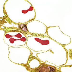
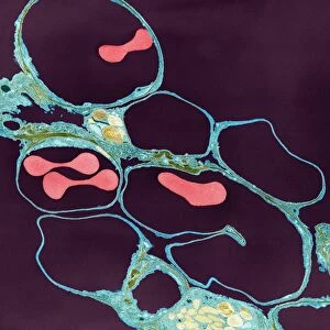
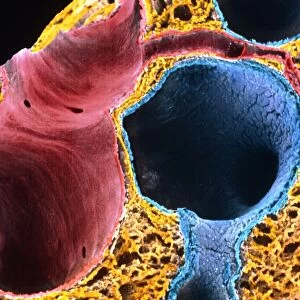
Lung lining, SEM
Lung lining. Coloured scanning electron micrograph (SEM) of mucus-producing cells (orange, round) and cilia (yellow) lining a bronchus (lung airway). Mucus secreted here traps bacteria, dust and other particles. Rhythmic movements of the cilia serve to move the mucus and the trapped particles away from the gas-exchanging parts of the lung, and towards the throat, where they can be expelled
Science Photo Library features Science and Medical images including photos and illustrations
Media ID 6450441
© SUSUMU NISHINAGA/SCIENCE PHOTO LIBRARY
Airway Bronchus Cellular Cilia Ciliated Cilium Epithelial Epithelium Internal Lining Lung Mucosa Mucosal Mucous Cell Mucus Orange Physiological Physiology Respiratory Tissue Cells False Coloured
21.5"x17.5" (55x44cm) Premium Frame
Discover the intricacies of the human body with Media Storehouse's Framed Prints featuring "Lung lining, SEM" by Science Photo Library. This captivating coloured Scanning Electron Micrograph (SEM) showcases the intricate detail of mucus-producing cells and cilia lining a bronchus, offering a unique glimpse into the complex world of lung anatomy. Bring the beauty of science into your home or office with this striking and conversation-starting piece.
Framed and mounted 17x12 print. Professionally handmade full timber moulded frames are finished off with framers tape and come with a hanging solution on the back. Outer dimensions are 21.5x17.5 inches (546x444mm). Quality timber frame frame moulding (20mm wide and 30mm deep) with frame colours in your choice of black, white, or raw oak and a choice of black or white card mounts. Frames have a perspex front providing a virtually unbreakable glass-like finish which is easily cleaned with a damp cloth.
Contemporary Framed and Mounted Prints - Professionally Made and Ready to Hang
Estimated Image Size (if not cropped) is 41.8cm x 41.8cm (16.5" x 16.5")
Estimated Product Size is 44.4cm x 54.6cm (17.5" x 21.5")
These are individually made so all sizes are approximate
Artwork printed orientated as per the preview above, with landscape (horizontal) or portrait (vertical) orientation to match the source image.
EDITORS COMMENTS
This print from Science Photo Library showcases the intricate beauty of lung lining at a microscopic level. In this false-colored scanning electron micrograph (SEM), we are presented with a mesmerizing display of orange mucus-producing cells and yellow cilia that line a bronchus, one of the airways in our lungs. The mucus-producing cells, depicted as vibrant orange spheres, play a vital role in our respiratory system. They secrete mucus which acts as a sticky trap for bacteria, dust particles, and other harmful substances present in the air we breathe. Alongside them, the delicate yellow cilia can be seen protruding like tiny hairs. These cilia possess rhythmic movements that serve to propel the mucus and trapped particles away from the gas-exchanging regions of our lungs towards our throat. Through this efficient mechanism, these remarkable structures ensure that any potential threats or irritants are expelled from our bodies before they reach deeper into our respiratory system. This image provides us with an awe-inspiring glimpse into the internal workings of our lungs and highlights their incredible ability to protect us against external pollutants. Science Photo Library has once again captured nature's marvels through their lens, reminding us of both the complexity and elegance found within even the tiniest components of life.
MADE IN AUSTRALIA
Safe Shipping with 30 Day Money Back Guarantee
FREE PERSONALISATION*
We are proud to offer a range of customisation features including Personalised Captions, Color Filters and Picture Zoom Tools
SECURE PAYMENTS
We happily accept a wide range of payment options so you can pay for the things you need in the way that is most convenient for you
* Options may vary by product and licensing agreement. Zoomed Pictures can be adjusted in the Cart.