Fine Art Print > Arts > Minimalist artwork > Monochrome artwork > Fine art
Fine Art Print : Liver tissue, TEM


![]()
Fine Art Prints from Science Photo Library
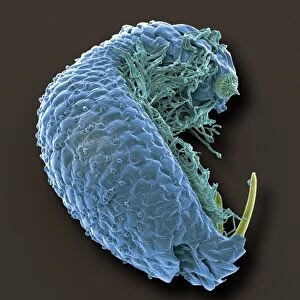
Liver tissue, TEM
Liver tissue. Transmission electron micrograph (TEM) of a section through the liver, showing part of a radial cord of hepatocyte liver cells (dark) and the vascular sinusoids (white). The hepatocyte cytoplasm notably contains mitochondria, rough endoplasmic reticulum and large amounts of glycogen. Sinusoids are a type of capillary, a small blood vessel. In the liver, this network of vessels infiltrates the tissue, supplying it with blood. Gases and nutrients are exchanged between the blood and surrounding tissue through the permeable walls of the capillaries. The gap between hepatocytes and the endothelium, known as the space of Disse, contains sparse collagen (reticular) fibres. Magnification: x2, 500 when printed 10 centimetres tall
Science Photo Library features Science and Medical images including photos and illustrations
Media ID 9240693
© MICROSCAPE/SCIENCE PHOTO LIBRARY
Black And White Capillaries Capillary Cell Biology Collagen Cytological Cytology Cytoplasm Endoplasmic Reticulum Glycogen Granule Granules Hepatic Hepatocyte Hepatocytes Histological Histology Liver Metabolic Metabolism Mitochondria Mitochondrion Network Organelle Organelles Sinusoids Transmission Electron Micrograph Transmission Electron Microscope Vascular System Vessels Blood Vessel Cells Circulation Circulatory System Section Sectioned
20"x16" (+3" Border) Fine Art Print
Discover the intricacies of life with our Fine Art Prints from Media Storehouse. This captivating image, captured through Transmission Electron Microscopy by Science Photo Library, offers a mesmerizing glimpse into the complex structure of the liver. Witness the intricate arrangement of hepatocyte liver cells and the vascular sinusoids in their true glory. A must-have for science enthusiasts and art connoisseurs alike, these prints bring the beauty of the microscopic world into your home or office space.
20x16 image printed on 26x22 Fine Art Rag Paper with 3" (76mm) white border. Our Fine Art Prints are printed on 300gsm 100% acid free, PH neutral paper with archival properties. This printing method is used by museums and art collections to exhibit photographs and art reproductions.
Our fine art prints are high-quality prints made using a paper called Photo Rag. This 100% cotton rag fibre paper is known for its exceptional image sharpness, rich colors, and high level of detail, making it a popular choice for professional photographers and artists. Photo rag paper is our clear recommendation for a fine art paper print. If you can afford to spend more on a higher quality paper, then Photo Rag is our clear recommendation for a fine art paper print.
Estimated Image Size (if not cropped) is 40.6cm x 50.8cm (16" x 20")
Estimated Product Size is 55.9cm x 66cm (22" x 26")
These are individually made so all sizes are approximate
Artwork printed orientated as per the preview above, with portrait (vertical) orientation to match the source image.
FEATURES IN THESE COLLECTIONS
> Arts
> Minimalist artwork
> Monochrome artwork
> Fine art
> Arts
> Minimalist artwork
> Monochrome artwork
> Monochrome paintings
EDITORS COMMENTS
This print showcases the intricate structure of liver tissue at a microscopic level. Taken using a transmission electron microscope (TEM), the image reveals a section through the liver, highlighting both hepatocyte liver cells and vascular sinusoids. The radial cord of dark hepatocyte cells is prominently displayed, surrounded by the white network of vascular sinusoids. These sinusoids are specialized capillaries that infiltrate the liver tissue, ensuring its blood supply. Through their permeable walls, gases and nutrients are exchanged between the blood and surrounding tissues. The hepatocytes themselves exhibit remarkable features within their cytoplasm. Abundant mitochondria can be observed alongside rough endoplasmic reticulum, indicating high metabolic activity in these cells. Additionally, large amounts of glycogen granules are present, serving as an energy reserve for various cellular processes. Notably, there is sparse collagen fiber content in the space known as Disse's space - this gap separates hepatocytes from endothelial cells lining the sinusoids. With a magnification factor of x2,500 when printed at 10 centimeters tall, this photograph provides an awe-inspiring glimpse into the complex biology and circulatory system found within our livers. It serves as a testament to how advanced imaging techniques can unravel hidden details within our bodies' tissues and organs.
MADE IN AUSTRALIA
Safe Shipping with 30 Day Money Back Guarantee
FREE PERSONALISATION*
We are proud to offer a range of customisation features including Personalised Captions, Color Filters and Picture Zoom Tools
SECURE PAYMENTS
We happily accept a wide range of payment options so you can pay for the things you need in the way that is most convenient for you
* Options may vary by product and licensing agreement. Zoomed Pictures can be adjusted in the Cart.